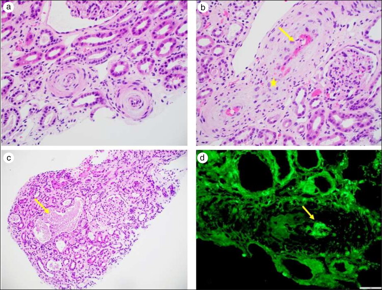
Figure 2.
Renal biopsy. (a) Arterioles with concentric intimal fibrosis and mucoid changes with swollen endothelial cells resulting in near luminal occlusion (light microscopy, hematoxylin-eosin stain, 400× magnification). (b) Interlobar artery with fibrin deposition (arrow) and fragmented red blood cells (star) in the vascular wall (light microscopy, hematoxylin-eosin stain, 400× magnification). (c) Tubular necrosis with sloughing of the tubular epithelial cells (arrow) (light microscopy, hematoxylin-eosin stain, 200× magnification). (d) Immunofluorescence microscopy with fibrinogen highlighting the presence of intravascular fibrin thrombus (immunofluorescence microscopy, fibrinogen, 400× magnification).

