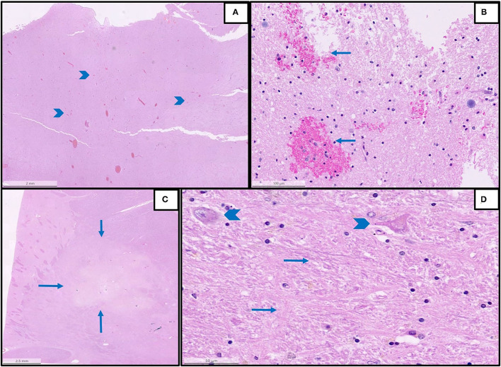
Figure 2.
Representative histopathological findings in autopsies from the first wave. (A) An overview of multifocal intracerebral microhemorrhages (arrowheads) in a sample from the brain stem (H&E, magnification 1.2 x). (B) The detail of image A with microhemorrhages (magnification 25 x, arrows: microhemorrhages). (C) An overview of acute brain infarction (arrows) in a sample from basal ganglia (H&E, magnification.62 x). (D) Details of image C showing red neurons (arrowheads) and beginning necrosis (arrows) of brain tissue (H&E, magnification 40 x).

