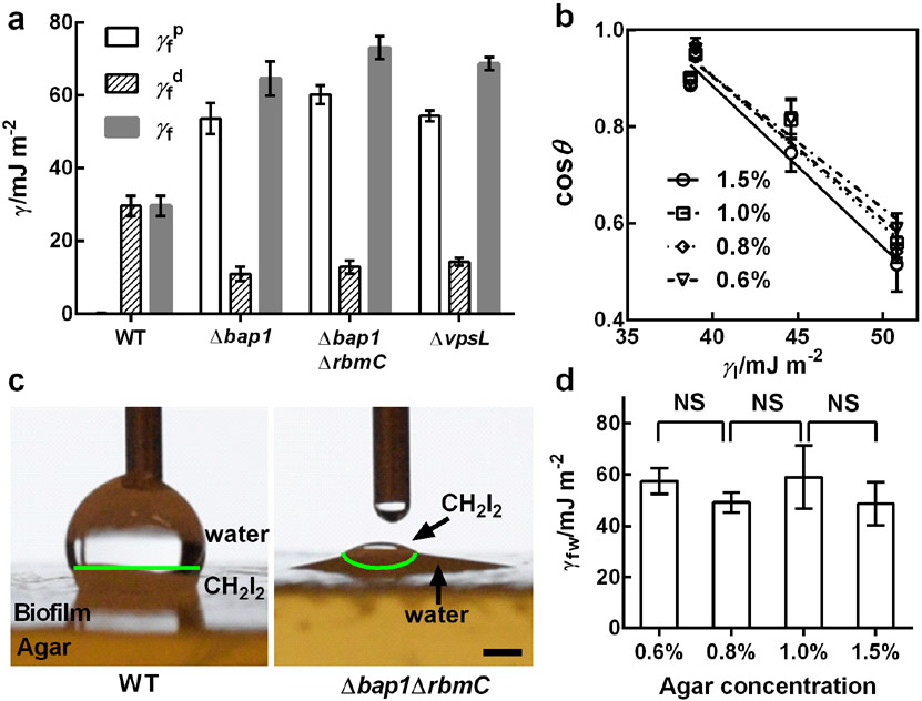
Figure 3.
Characterization of biofilm surface energies. a) Measured values of the polar () and dispersion () components of the surface energy (γf) for the designated V. cholera biofilms grown for two days on 1.5% agar using the Owens-Wendt method.[39] b) Measurement of the surface energies of WT biofilms grown on medium containing the designated concentrations of agar, using the Zisman method.[40] The contact angles θ for nonpolar liquids (CH2I2, CH2Br2, 1-methylnaphthalene, and 1-bromonaphthalene) on biofilms were measured and plotted against the surface energies of the liquids (γ1) for different concentrations of agar. Linear extrapolation to cosθ = 1 gives the critical surface tension for wetting γc corresponding to the nonpolar component of the surface energy. The symbols designate measurements made for biofilms grown on LB medium solidified with the designated agar concentrations. c) Representative images showing preferential wetting of the WT biofilm by a polar (water) liquid and the Δbap1ΔrbmC mutant biofilm by a nonpolar (CH2I2) liquid, when both liquids were present on top of each biofilm. The green lines denote the interfaces between water and CH2I2. Scale bar: 1 mm. d) Measured biofilm-water interfacial energies γfw for biofilms grown on medium solidified with the designated concentrations of agar. Unpaired t-tests with Welch’s correction were performed for statistical analyses. NS stands for not significant. Error bars correspond to standard deviations with n = 3.

