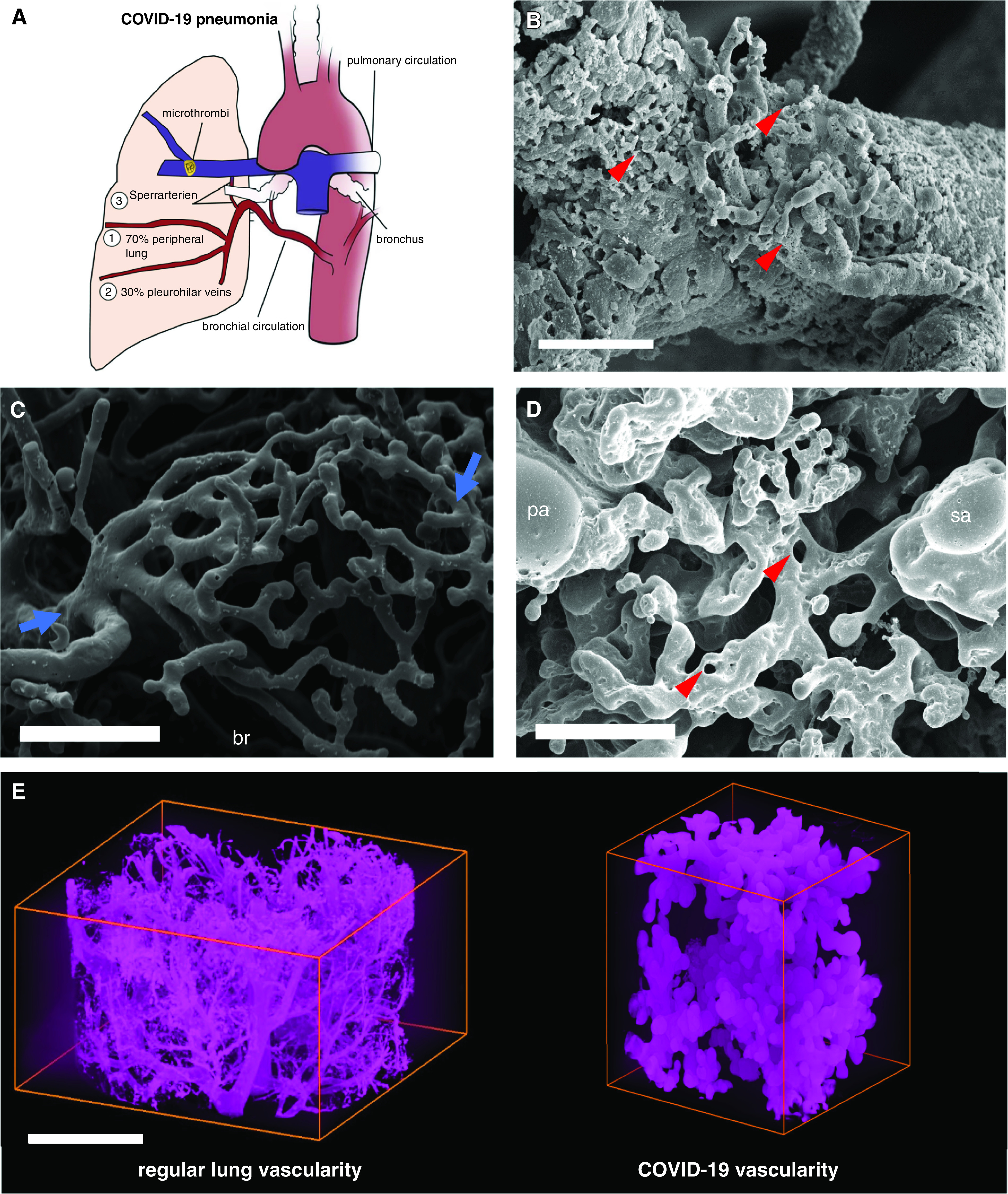
Figure 2.

Intrapulmonary shunting via bronchial circulation in coronavirus disease (COVID-19) pneumonia. (A) The schematic illustrates intrapulmonary cross-circulation and draining by bronchial arteries to the peripheral lung, pleurohilar veins, and “Sperrarterien,” specialized, spiral-like intrapulmonary arteriovenous anastomoses recruited by hypoxia and flow irregularities. The bronchial circulation arises primarily direct from the thoracic aorta and intercostal arteries and, under normal conditions, constitutes less than 2% volume of the left cardiac output. However, the bronchial flow can significantly exceed the regular hemodynamics upon hypoxic and inflammatory stimulus. As part of a physiological right–left shunting, nearly two-thirds of the bronchial vessels are drained peribronchially to the peripheral lung and finally into the left ventricle (up to 4% of left ventricular output), whereas the remaining third is perfused pleurohilar veins draining ultimately to the right heart. (B) Scanning electron micrograph of a microvascular corrosion cast demonstrates the expansion of vasa vasorum of an intralobular artery by intussusceptive angiogenesis (red arrowheads) in the lung tissue of a patient who succumbed to severe COVID-19 pneumonia. Scale bar, 50 μm. Intussusceptive angiogenesis is a well-characterized morphogenetic process observed during the capillary expansion in cancer, inflammation, and regeneration, but it has also been reported in the alveolar plexus of COVID-19 lungs. Scale bar, 100 μm. (C) Scanning electron micrograph of a microvascular corrosion cast demonstrates the peribronchial plexus in healthy control lungs (blue arrows). Scale bar, 200 μm. (D) Scanning electron micrograph of a microvascular corrosion cast reveals the cross-circulation in COVID-19 lung tissue between a Sperrarterie and a lobular artery in a secondary pulmonary lobule, which is characterized by plexiform-like vascular proliferations by intussusceptive angiogenesis (red arrowheads). Scale bar, 200 μm. (E) Three-dimensional evaluation of microvascular corrosion casts by Synchrotron radiation X-ray tomographic microscopy illustrating the irregular, hierarchical vascularity with dilated, blunt-like vessels in COVID-19 pneumonia. Scale bar, 100 μm. br = bronchiole, pa = lobular artery; sa = Sperrarterie.
