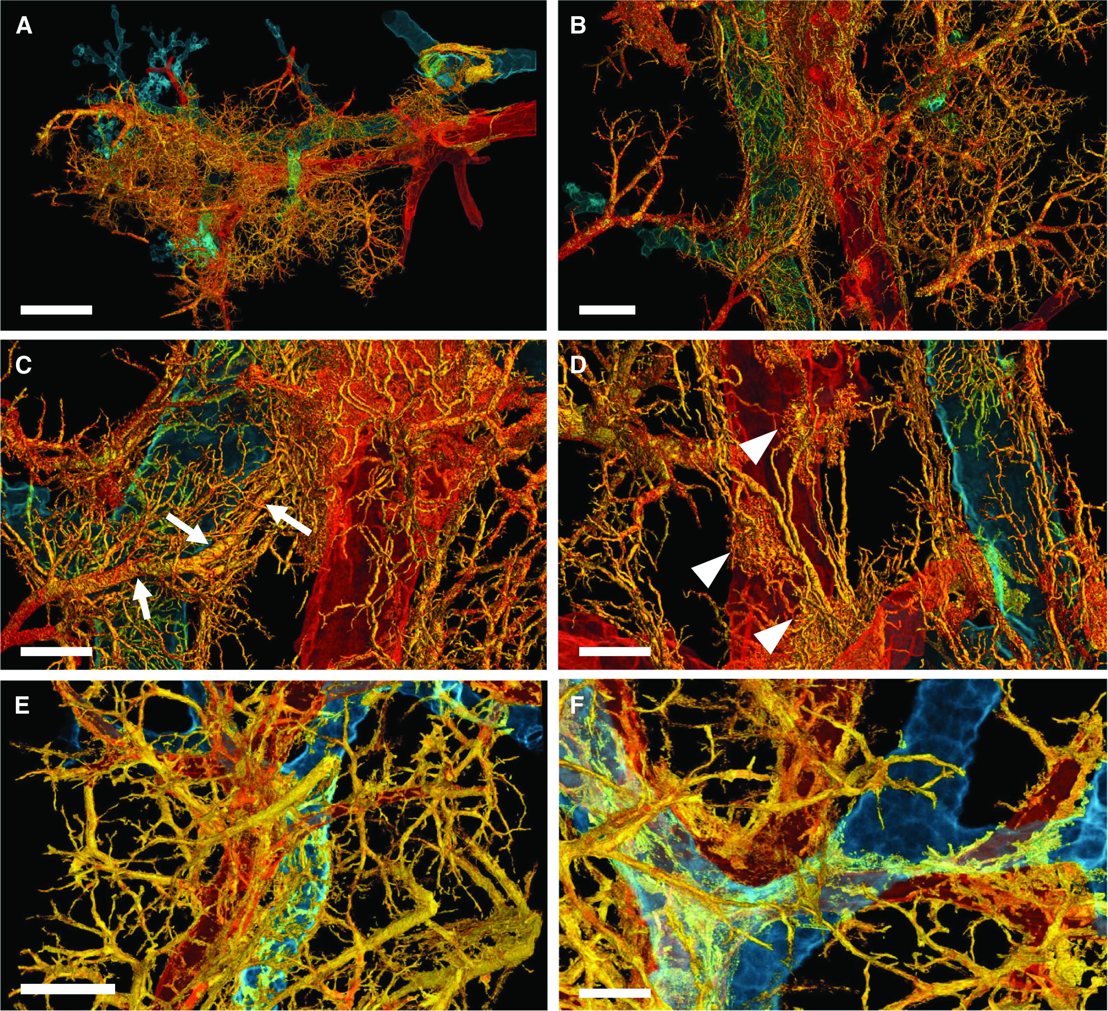
Figure 3.

Three-dimensional complexity of vascular remodeling and bronchiopulmonary shunting in coronavirus disease (COVID-19) highlighted by hierarchical phase-contrast tomography. (A) The distal bronchiole (blue) is paralleled by a centrilobular pulmonary artery (red) and an expanded plexus of private vessels (vasa vasorum) (yellow) in a secondary pulmonary lobule in the periphery of the left lower lobe. Scale bar, 4 mm. (B) Dilatation and expansion of the peribronchiolar plexus and vasa vasorum (yellow) is observed around the distal terminal bronchiole (blue) and centrilobular pulmonary artery (red). Scale bar, 1.3 mm. (C) Numerous arteriovenous anastomoses (yellow) form between bronchial arteries and pulmonary artery drain into a pulmonary vein accompanied by a pronounced shunting of a Sperrarterie (white arrows). These specialized arteries are essential for intrapulmonary arteriovenous shunting and are located mainly at the septal margin of secondary pulmonary lobules, where up to 3 to 5 such “Sperrarterien” anastomoses can be found in each lobule. Scale bar, 0.7 mm. (D) The perivascular vasa vasorum and bronchial vascular plexus are characterized by complex nodular lesions with angiomatoid- and plexiform-like arrangements (white arrowheads). Scale bar, 0.7 mm. For high-resolution details, see COVID-19 Pneumonia in the online supplement . (E) In controls, the vascular architecture shows a well-organized hierarchy of peribronchial vessels. Scale bar, 1.3 mm. (F) Only few perivascular vasa vasorum are observed in healthy control tissue. Scale bar, 0.7 mm. For high-resolution details, see Control in the online supplement.
