Figure 1.

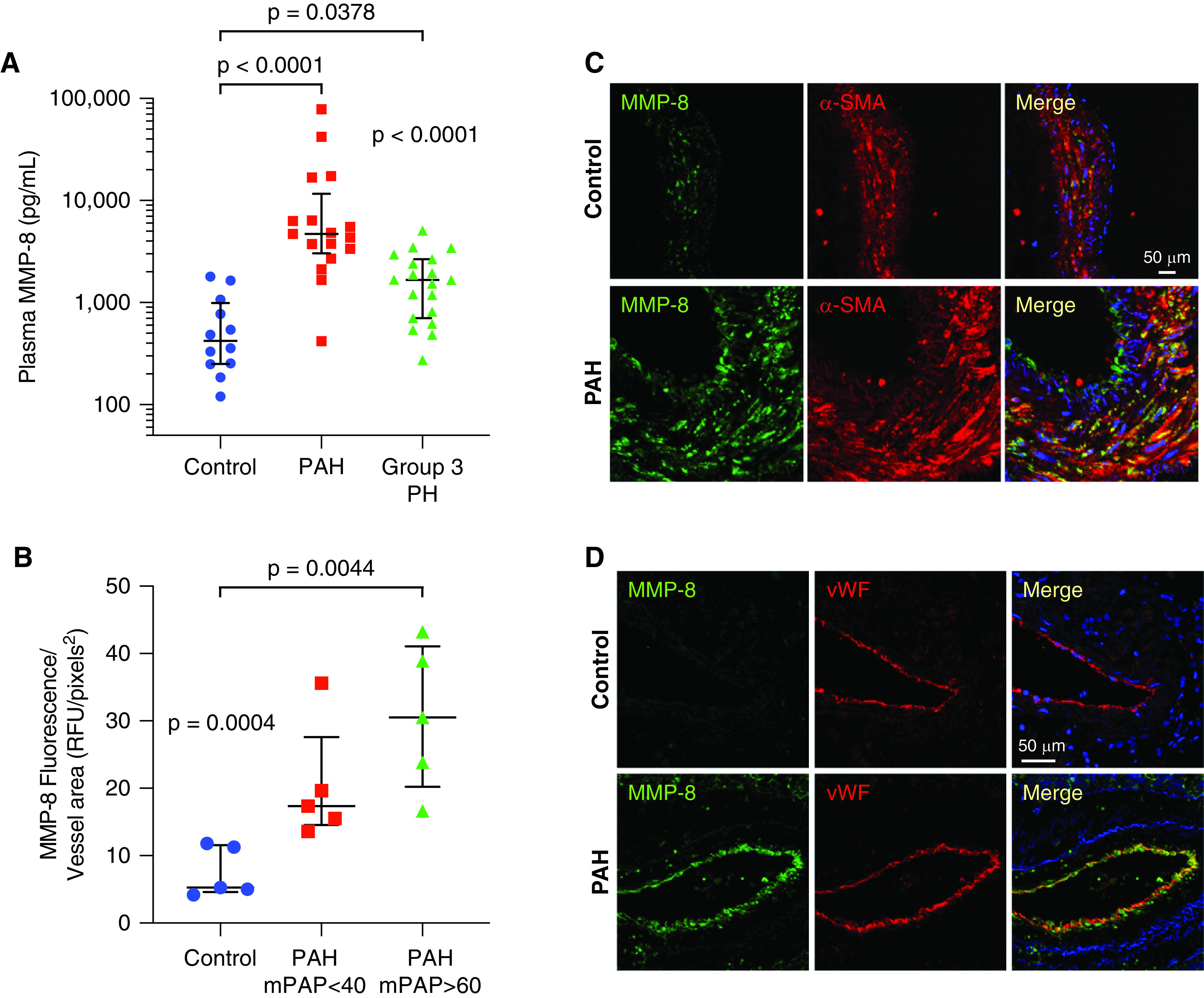
MMP-8 (matrix metalloproteinase-8) expression is increased in plasma and pulmonary arteries (PAs) of patients with pulmonary arterial hypertension (PAH). (A) MMP-8 levels were measured in plasma from patients with group 1 PAH (n = 17, 64 ± 14 yr), patients with group 3 pulmonary hypertension (PH) (n = 19, 65 ± 8 yr), and age-matched controls without PH (n = 12, 66 ± 8 yr) by ELISA. Data represent median and interquartile range (IQR). Statistical significance was determined by Kruskal-Wallis one-way ANOVA (P < 0.0001) followed by Dunn’s multiple comparisons test (P < 0.0001 group 1 PAH vs. controls, P = 0.0378 group 3 PH vs. controls). (B–D) Immunofluorescence staining for MMP-8, α-SMA (α-smooth muscle actin), and vWF (von Willebrand factor) was performed in explanted lungs from patients with group 1 PAH (mPAP < 40 mm Hg, mPAP > 60 mm Hg) who underwent lung transplantation and control lungs (n = 5 per group). (B) MMP-8 fluorescence intensity was measured in 10 PAs (50–150 μm) per patient and normalized per vessel area using Metamorph software. The mean MMP-8 fluorescence intensity per vessel area for each patient is shown with median and IQR per group. Statistical significance was determined by Kruskal-Wallis one-way ANOVA (P = 0.0004) followed by Dunn’s multiple comparisons test (P = 0.0044 control vs. PAH with mPAP > 60 mm Hg). (C and D) Representative confocal images are shown for MMP-8 (green) and α-SMA (red) immunostaining (C) and MMP-8 (green) and vWF (red) immunostaining (D). Merged images are shown on the right of each panel. Scale bars, 50 μm. mPAP = mean pulmonary arterial pressure; RFU = relative fluorescence units.
