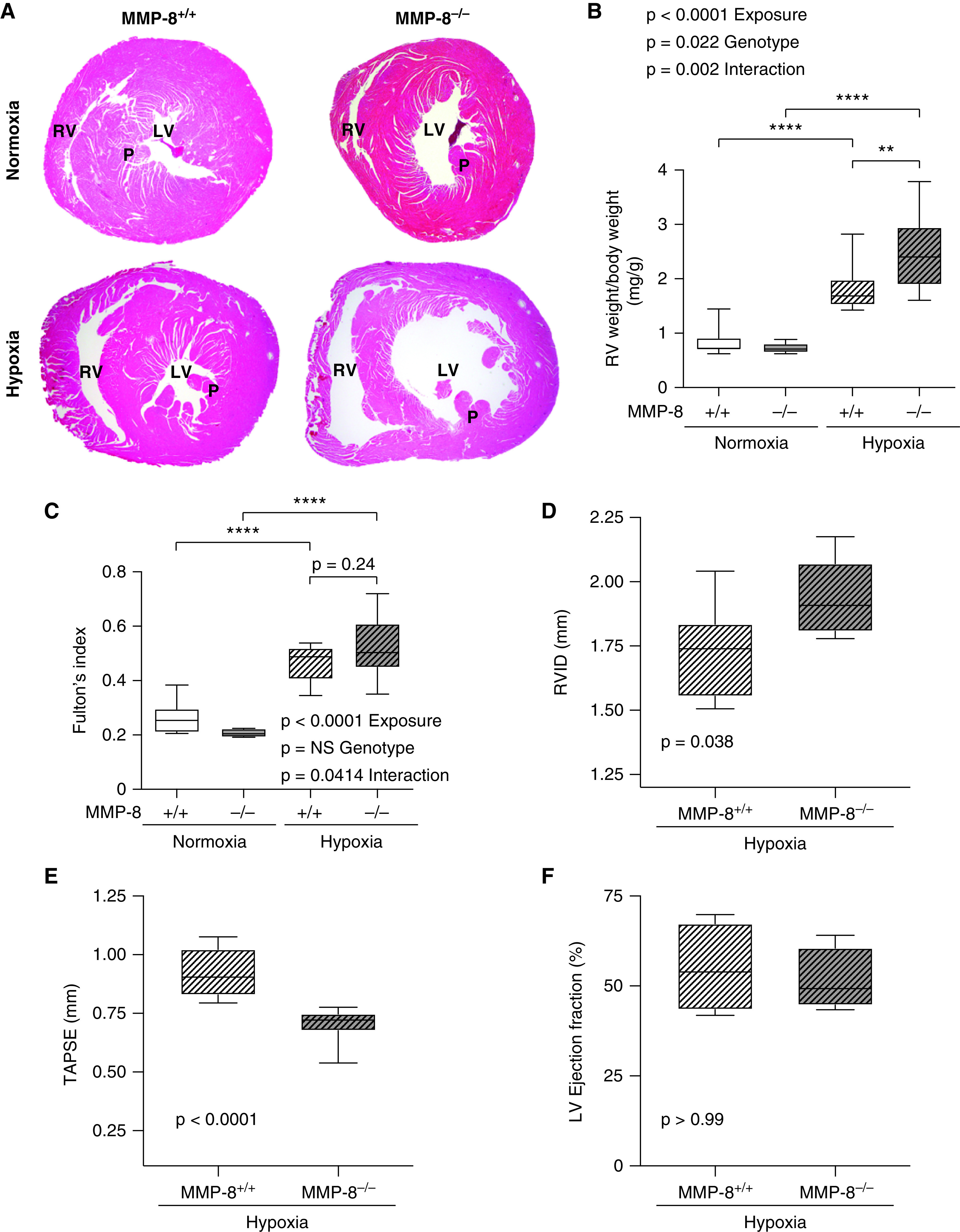
Figure 4.

MMP-8 (matrix metalloproteinase-8) deficiency leads to RV hypertrophy and impairs RV function after chronic hypoxia. MMP-8+/+ and MMP-8–/– mice were exposed to 8 weeks of normobaric hypoxia or normoxia (n = 10–15 per group). (A) Representative cross-section images of hematoxylin and eosin–stained hearts at the level of the papillary muscles (P). (B and C) RV hypertrophy was assessed by normalizing RV weight (in milligrams) to total body weight (in grams) (B) and by assessing Fulton’s index (C). Data represent 25th–75th percentiles (box), median (line), and minimum and maximum values (whiskers). Statistical significance was determined by two-way ANOVA followed by Tukey’s post hoc test (**P ⩽ 0.01 and ****P ⩽ 0.0001). (D–F) Echocardiography was performed, and measurements of RV internal diameter (RVID), tricuspid annulus plane systolic excursion (TAPSE), and left ventricular ejection fraction were made in MMP-8+/+ and MMP-8–/– mice after 8 weeks of hypoxia (n = 8 per group). Data represent 25th–75th percentiles (box), median (line), and minimum and maximum values (whiskers). Statistical significance was determined by the Mann-Whitney U test. There was no significant difference in left ventricular ejection fraction between MMP-8+/+ and MMP-8–/– mice after hypoxia. LV = left ventricle; NS = not significant; RV = right ventricle.
