Figure 7.

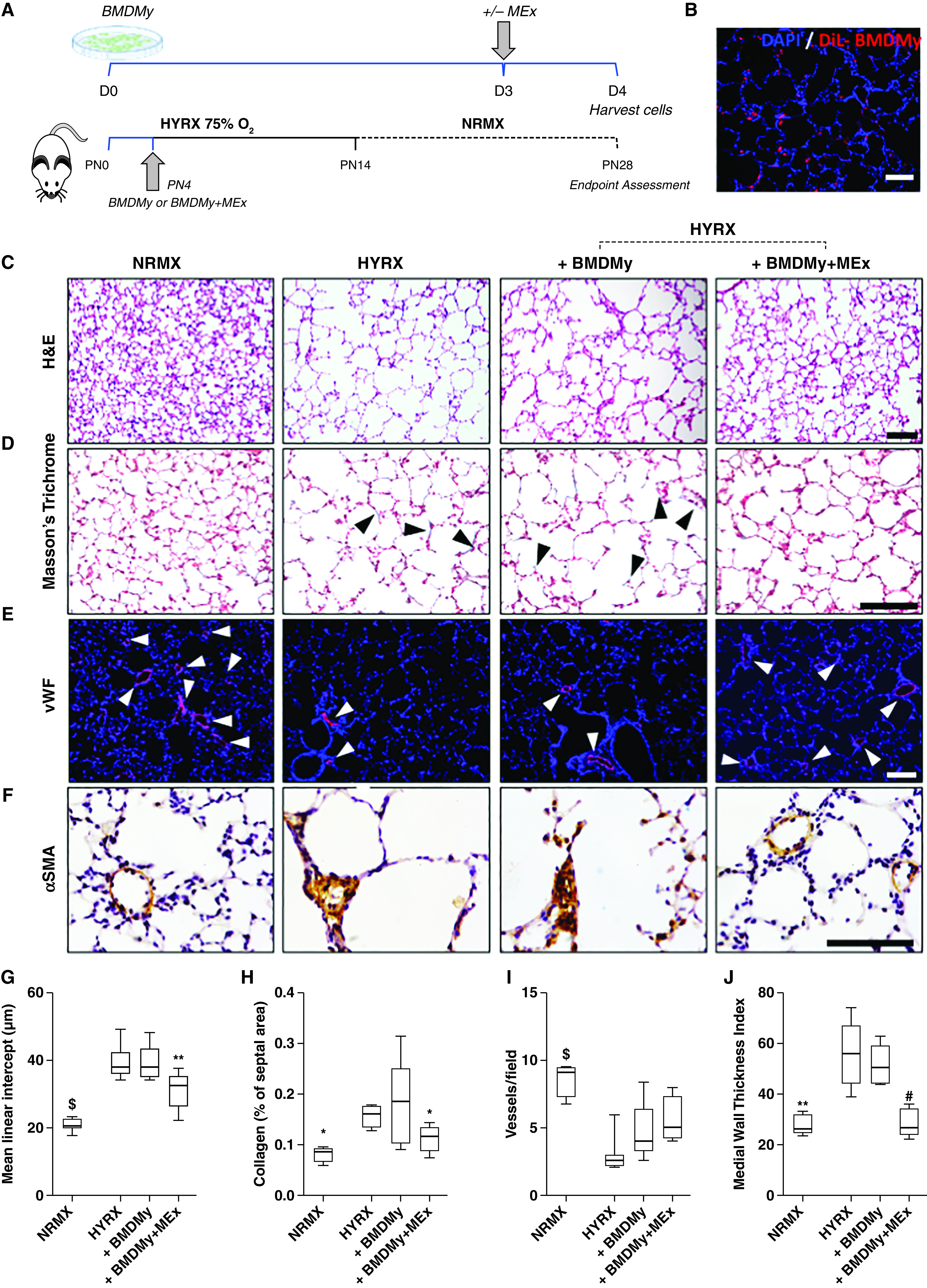
Adoptive transfer of mesenchymal stem/stromal cell–small extracellular vesicle (MEx)-educated bone marrow-derived myeloid cells (BMDMy) increases alveolarization and reduces pulmonary fibrosis and hyperoxia (HYRX)-induced vascular muscularization. (A) Schematic representation of adoptive transfer workflow. Briefly, newborn Friend leukemia virus B mice were exposed to HYRX (75% O2) for 14 days. HYRX-exposed mice were compared with mice that remained at room air (normoxia [NRMX]). BMDMy or MEx-educated BMDMy were delivered intravenously at PN4. Animals were assessed at PN28. (B) Proof-of-concept studies showed that dialkylcarbocyanine (DiL)-labeled BMDMy were detected in the lung at PN14. Blue = DAPI. (C–F) Harvested lung sections were stained for hematoxylin and eosin (H&E) to assess lung architecture (C), Masson’s trichrome to evaluate septal collagen deposition (D), and vWF (von Willebrand factor) (E) or α-SMA (α-smooth muscle actin) (F) to assess the effect of HYRX on peripheral pulmonary blood vessel loss and pulmonary vascular remodeling, respectively. (G–J) Quantification of mean linear intercept (μm) (G), collagen deposition (reported as % of septal area) (H), blood vessel count (positive <500-μm outer diameter) (I), and medial thickness index (J). Black arrowheads highlight collagen deposition. White arrowheads highlight vWF-stained pulmonary vessels. Data represent results from three individual studies. n = 6–12 per group; *P < 0.05, **P < 0.01, #P < 0.001, and $P < 0.0001 versus HYRX. Scale bars: H&E and vWF, 100 μm; Masson’s trichrome and α-SMA, 50 μm. PN = Postnatal Day.
