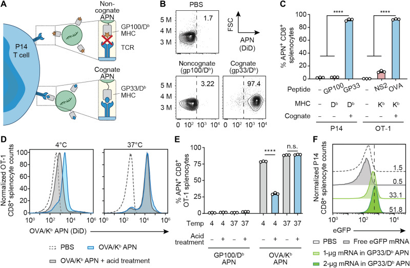
Fig. 2. APNs target Ag-specific T cells and induce cell uptake in vitro.
(A) Illustration of the interaction of P14 CD8+ T cells with its cognate APNs (GP33/Db), in contrast to the lack of binding to the noncognate control (GP100/Db APN). (B) Representative flow plots of noncognate APNs and cognate APNs binding to CD8+ T cells in splenocytes from P14 TCR transgenic mice. Frequencies depicted are based on gating on CD8+ cells. (C) Ag-specific binding of cognate and noncognate APNs to CD8+ T cells isolated from TCR transgenic P14 or OT-1 mice. ****P < 0.0001, one-way analysis of variance (ANOVA) and Tukey post-test and correction. All data are means ± SD; n = 3 biologically independent wells. (D and E) OT-1 CD8+ splenocytes stained with OVA/Kb APNs at 4° or 37°C and analyzed by flow cytometry before and after treatment with an acetate buffer to strip cell surface proteins. GP100/Db APNs served as a noncognate control. ****P < 0.0001 between OVA/Kb APN treatment with and without acid treatment at 4°C; n.s., not significant, where P = 0.61 between OVA/Kb APN with and without acid treatment at 37°C; two-way ANOVA and Sidak post-test and correction. All data are means ± SD; n = 3 biologically independent wells. (F) eGFP mRNA expression in P14 CD8+ T cells after coincubation with free-form eGFP mRNA or eGFP mRNA loaded in GP33/Db APNs for 24 hours.

