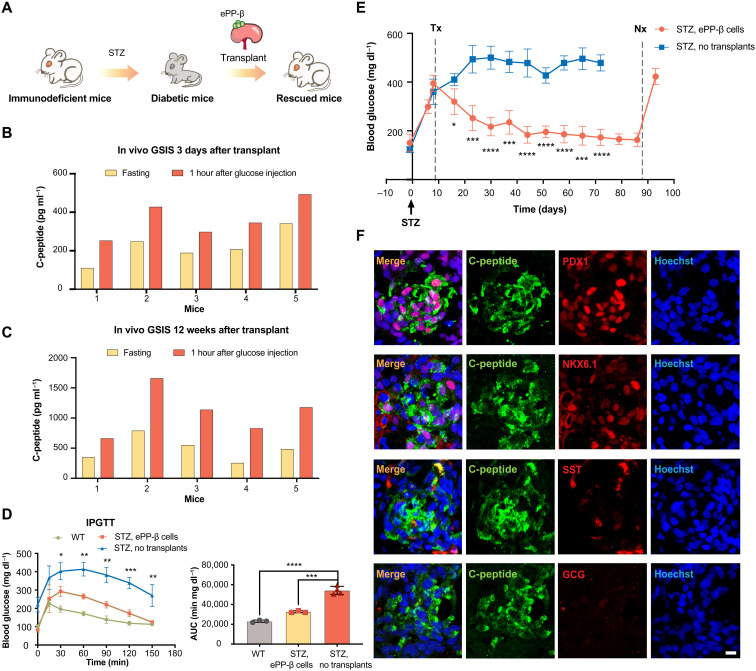
Fig. 4. ePP-β cells can rapidly ameliorate diabetes.
(A) Schematic showing transplantation of ePP-β cells into immunodeficient diabetic mice. (B and C) In vivo glucose challenge test 3 days (B) and 12 weeks (C) after transplantation. The levels of human C-peptide in mouse serum were measured after overnight fasting (yellow) and 1 hour after an intraperitoneal glucose injection (orange). The x axis represents the number of individual animals (N = 5). (D) Intraperitoneal glucose tolerance test (IPGTT) was performed 30 days after transplantation with ePP-β cells (N = 3), nontransplanted diabetic SCID beige mice (N = 4), and nontransplanted wild-type (WT) SCID beige mice (N = 3). Area under the curve (AUC; left) was determined for each group (right). (E) Blood glucose level of STZ-treated control mice (no transplant, blue) and mice transplanted with ePP-β cells (orange). Tx, transplantation; Nx, nephrectomy. Control group (N = 5) and experimental group (N = 4). (F) Representative immunofluorescent images of ePP-β grafts stained with C-peptide, NKX6.1, PDX1, SST, GCG, and nuclei. Scale bar, 10 μm. All data are expressed as means ± SD. Statistical significance was calculated using two-tailed Student’s t test, *P < 0.05, **P < 0.01, ***P < 0.001, and ****P < 0.0001.

