
Figure 1: OCT Images of Heart Defects Induced by Ethanol Exposure.
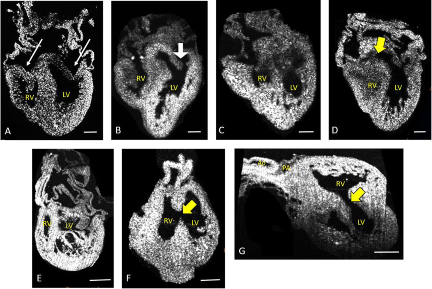
All images are of 8 day, HH stage 34 hearts. Panel (A) is of a normal heart, with long arrows pointing to normal left and right atrioventricular (AV) valve leaflets. Remaining panels show representative abnormalities: (B) Hypoplastic right ventricle and abnormal left AV valve leaflets (large white arrow), (C) Severely hypoplastic right ventricle, (D) ventricular septal defect, (E) left ventricular noncompaction, (F) and (G) different views of the same heart with double outlet right ventricle and large ventricular spetal defect. Yellow arrows point to ventricular septal defects. All scale bars are 50µm. Not all images are standard 4 chamber views, as that view cannot show all defects. RV = right ventricle, LV = Left Ventricle, PA = Pulmonary Artery, Ao = Aorta