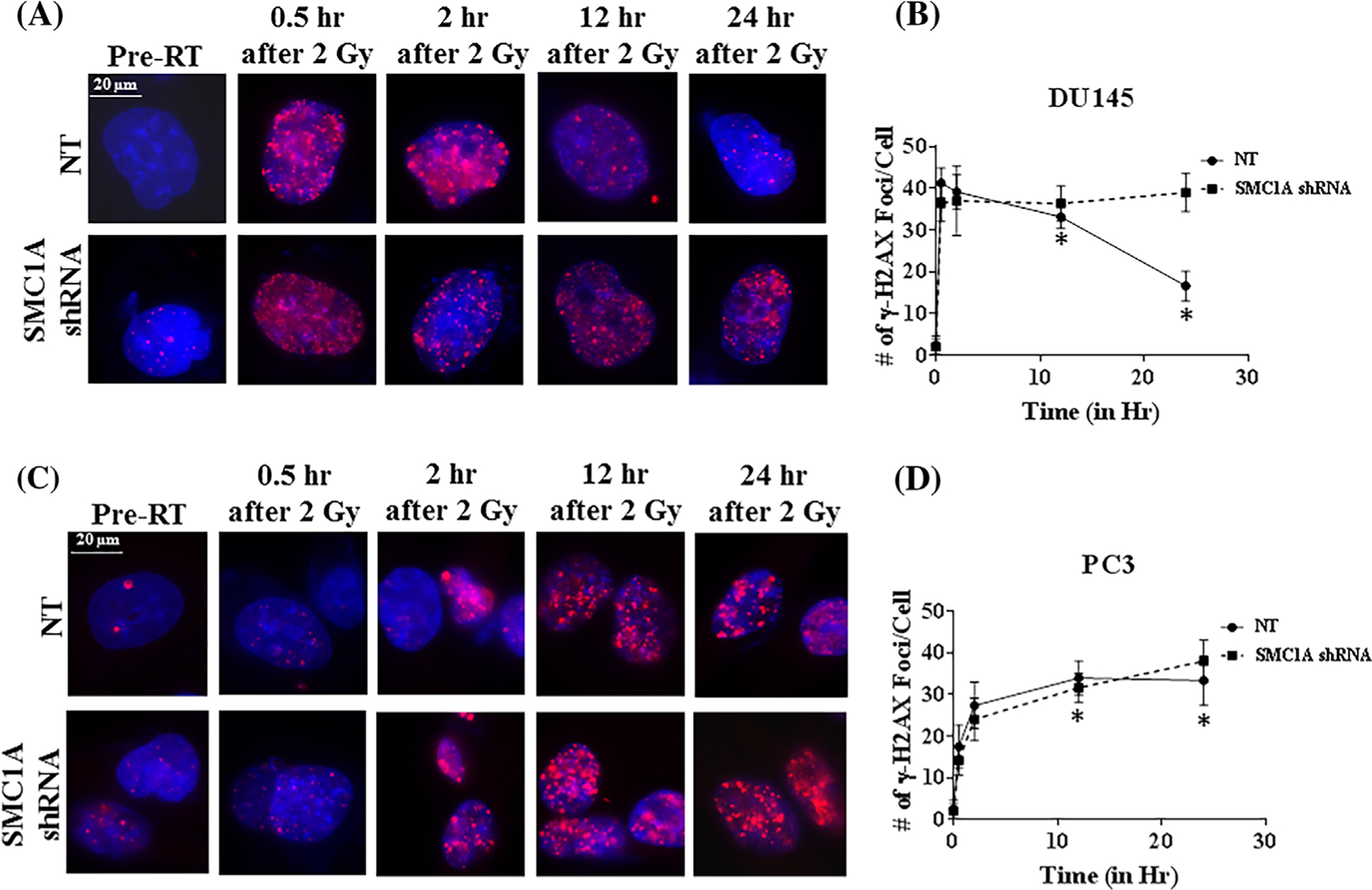
FIGURE 7.

Quantification of DNA DSB marker, γH2AX foci after irradiation in SMC1A shRNA expressing DU145 and PC3 cells. A, Representative images of γH2AX staining at the specific time points in NT and SMC1A-shRNA expressing cells after irradiation (2 Gy). Red fluorescence staining indicates positive while blue stain from DAPI indicates nuclei (magnification 40×). B, Number of γH2AX foci in SMC1A shRNA expressing DU145 and PC3 cells (Mean ± SD, n = 20) were counted and plotted. At 12 and 24 h after radiation (asterisks), significant difference was found between SMC1A shRNA expressing DU145 cells. The difference was more pronounced in DU145 cells compared to PC3 cells showing these cells are more radio-resistant. SMC1A suppression sensitized both DU145 and PC3 cells (P < 0.05; SMC1A shRNA vs NT shRNA expressing DU145 and PC3 cells, no radiation treatment and irradiated with single dose of 2 Gy)
