. 2022 Feb 23;11:e69504. doi: 10.7554/eLife.69504

Figure 1. Principle of the prime editor activity reporter (PEAR) assay.

(A) Schematic of the PEAR. The mechanism of PEAR is based on the same concept as BEAR (see Figure 1—figure supplement 1A and B), and it contains the same inactive splice site, shown in (A). PE can revert the ‘G-AC-AAGT’ sequence to the canonical ‘G-GT-AAGT’ splice site. Prime editing occurs downstream of the cut site in the target; hence, this method enables us to position the spacer sequence within the intron, thus, the entire length of the spacer (shown as ‘N’-s) is freely adjustable in PEAR. The altered bases of the splice site are shown in red, the edited bases are shown in blue, and the PAM sequence is shown as dark green letters, the nCas9 is blue and the fused reverse transcriptase is orange. (B) Optimization of PBS, RT, and complementary DNA strand nicking on the PEAR-GFP plasmid. The heatmap shows the average percentage of GFP-positive cells of three replicates of transfections with the PEAR-GFP plasmid in combination with PE vectors which also contain the different pegRNAs and the sgRNAs for secondary nicking. The position of the second nick is given in relation to the first nick. Positive values indicate 3′, negative values indicate 5′ direction on the targeted DNA. When no second nick was introduced, it is indicated as ‘no nick’. (C, D) Flow cytometry measurements of HEK293T cells transfected with active (positive control) or with inactive PEAR plasmids either along with nCas9 for negative controls or with PE2 vectors (black, gray, or green columns, respectively). (C) In the PEAR system, GFP fluorescence can be restored from various inactive splice sequences not only by substitutions but also by insertions/deletions. Each inactive sequence (gray columns) was corrected (green columns) to the canonical splice site by PE2 with the optimal pegRNA (pegRNA 1) from (B). The edited sequences are shown next to the columns. Red indicates inserted, blue substituted, and purple deleted bases. (D) Prime editing can result in various active splice sequences, further demonstrating the sequence flexibility of the PEAR system. Based on Tálas et al., 2021, four additional active splice site variants were selected. To generate suitable inactive plasmids, splicing was disrupted by systematically replacing the 5′-GT splice donor site to 5′-AC and 5′-CT (negative controls with inactive sequences, gray columns). With the appropriate pegRNAs, PE2 was able to restore GFP fluorescence in every case (green columns). Letters highlighted in blue indicate the bases of the canonical splice donor site and the flanking sequences which influence splicing the most: 5′-G-GT-AAGT-3′; the altered bases of the inactive sequences are bold, and bases in the active sequences that are reverted by PE2 are underlined. (C, D) Columns represent means ± SD of three parallel transfections (white circles). For all measured values see Figure 1—source data 1.

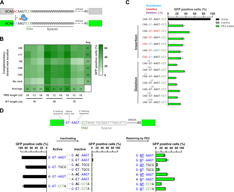
Figure 1—source data 1. Measured values for Figure 1.

Figure 1.

Figure 1—figure supplement 1. The differences between the base editor activity reporter (BEAR) and PEAR assays.

Figure 1—figure supplement 1.

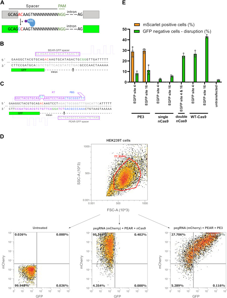
(A) Schematic representation of the BEAR. BEAR consists of a split GFP coding sequence separated by an intron, of which the 5′ splice donor site (G-GT-AAGT) is altered, resulting in an inactive splice site and a dysfunctional protein (gray). ABE converts the inactive splice site into a functional one. Here, the ‘G-AC-AAGT’ inactive splice site is illustrated, which can be modified by ABE to ‘G-GC-AAGT,’ which is a functional non-canonical splice site, and hence, restores GFP expression (green). In this assay, BEs act on the sense strand of the DNA. The altered bases of the splice site are shown in red, the edited base is shown in blue, and the variable bases in the sequence of the spacer are shown as ‘N’-s. The PAM sequence is dark green, nCas9 is blue, and the fused tadA deaminase is purple. (B) Detailed view of the 5′ splice site and the surrounding sequences of the BEAR-GFP plasmid. The 3′ end of the first exon of GFP is shown in green, the intron is shown as a dashed line. The spacer sequence and the target sequence are shown in gray, the PAM is green, and the inactive splice site is red. The Cas9 nick site is indicated by a black arrow. (C) Detailed view of the 5′ splice site and the surrounding sequences of the PEAR-GFP plasmid. The 3′ end of the first exon of GFP is shown in green, the intron is shown as a dashed line. The spacer sequence in the pegRNA and the target sequence in the DNA are shown in gray, the PAM is green, and the inactive splice site is red. The RT (purple) and the PBS (blue) sequences in the pegRNA and the targeted sequences are also colored. The Cas9 nick site is indicated by a black arrow. (D) A density plot of HEK293T cells when all live single cells (determined by FSC and SSC parameters) are either (1) untransfected (left plot), and thus show no fluorescence for mCherry or GFP, (2) co-transfected with a pegRNA-mCherry plasmid and the PEAR-GFP plasmid but with a nCas9, thus displaying mCherry but no GFP fluorescence, (3) or pegRNA-mCherry and PEAR-GFP plasmid is co-transfected with a prime editor expressing plasmid, causing the splice site to be edited, and thus displaying both mCherry and GFP fluorescence. (E) A HEK293T cell line containing an integrated EGFP expression cassette was co-transfected with the BEAR-mScarlet plasmid, a pegRNA and a second nicking sgRNA for BEAR-mScarlet, GFP targeting sgRNA(s) and either WT-Cas9, nCas9, or PE2. Cells were monitored for mScarlet and EGFP fluorescence (orange and green columns, respectively) 3 days after transfection. Columns represent means ± SD of three parallel transfections. For all measured values, see Figure 1—figure supplement 1—source data 1. PEAR, prime editor activity reporter.
Figure 1—figure supplement 1—source data 1. Measured values for Figure 1—figure supplement 1.