Figure 3.

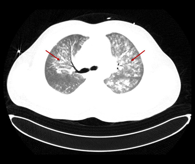
Chest computed tomography with contrast showing extensive bilateral ground-glass and patchy nodular opacities, more on the left lung (red arrows).

Chest computed tomography with contrast showing extensive bilateral ground-glass and patchy nodular opacities, more on the left lung (red arrows).