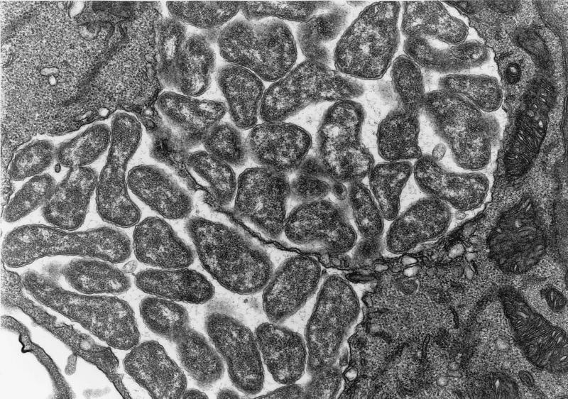
FIG. 3.
Electron micrograph of morulae of E. muris I-268 (A. speciosus, Tokyo isolate) in the cytoplasm of murine peritoneal cells at day 10 postinfection. Note the numerous pleomorphic coccobacilli enveloped in two layers of membranes embedded in a fine filamentous matrix in the membrane-bound inclusion. Magnification, ×22,100.

