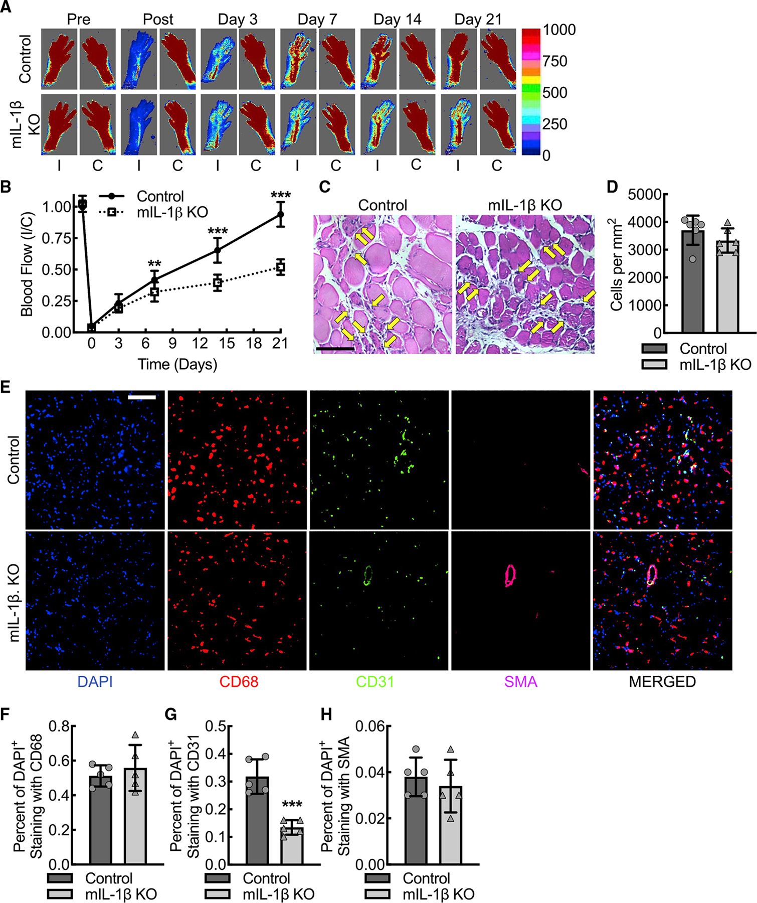
Figure 5. VEGF-A–induced blood flow recovery consequent to new angiogenesis and arteriogenesis is dependent on macrophage IL-1β expression in response to acute hindlimb ischemia.

(A and B) Laser Doppler images of flow in the ischemic (I) and contralateral control (C) hindlimbs of control or myeloid IL-1β-deleted mice (mIL-1β KO) at indicated time points before and after femoral artery ligation along with quantitative analysis (B) (**, p = 0.002; ***, p < 0.0001 between control and mIL-1β KO for each time point by ANOVA; n = 12 mice total, six males and six females).
(C and D) Standard histology stained for hematoxylin and eosin of day 3 ischemic gastrocnemius muscle tissue from control or mIL-1β KO mice along with quantification (D) of number of infiltrating inflammatory cells, indicated by yellow arrows (n = 6 mice total, three males and three females). Bar, 100 microns.
(E–H) Immunofluorescence micrographs of ischemic muscle tissue at day 3 post femoral artery ligation from control or mIL-1β KO mice along with quantitation of DAPI+CD68+ (F), DAPI+CD31+ (G), and DAPI+SMA+ (H) cells (***, p = 0.0003 by t test; n = 6 mice, three males and three females). Bar, 100 microns. Data, mean ± SD.
