
Figure 7. Clodronate liposome treatment followed by BMDM transplant in the context of acute hindlimb ischemia confirms the important contribution of IL-1β from macrophages relative to other leukocytes.
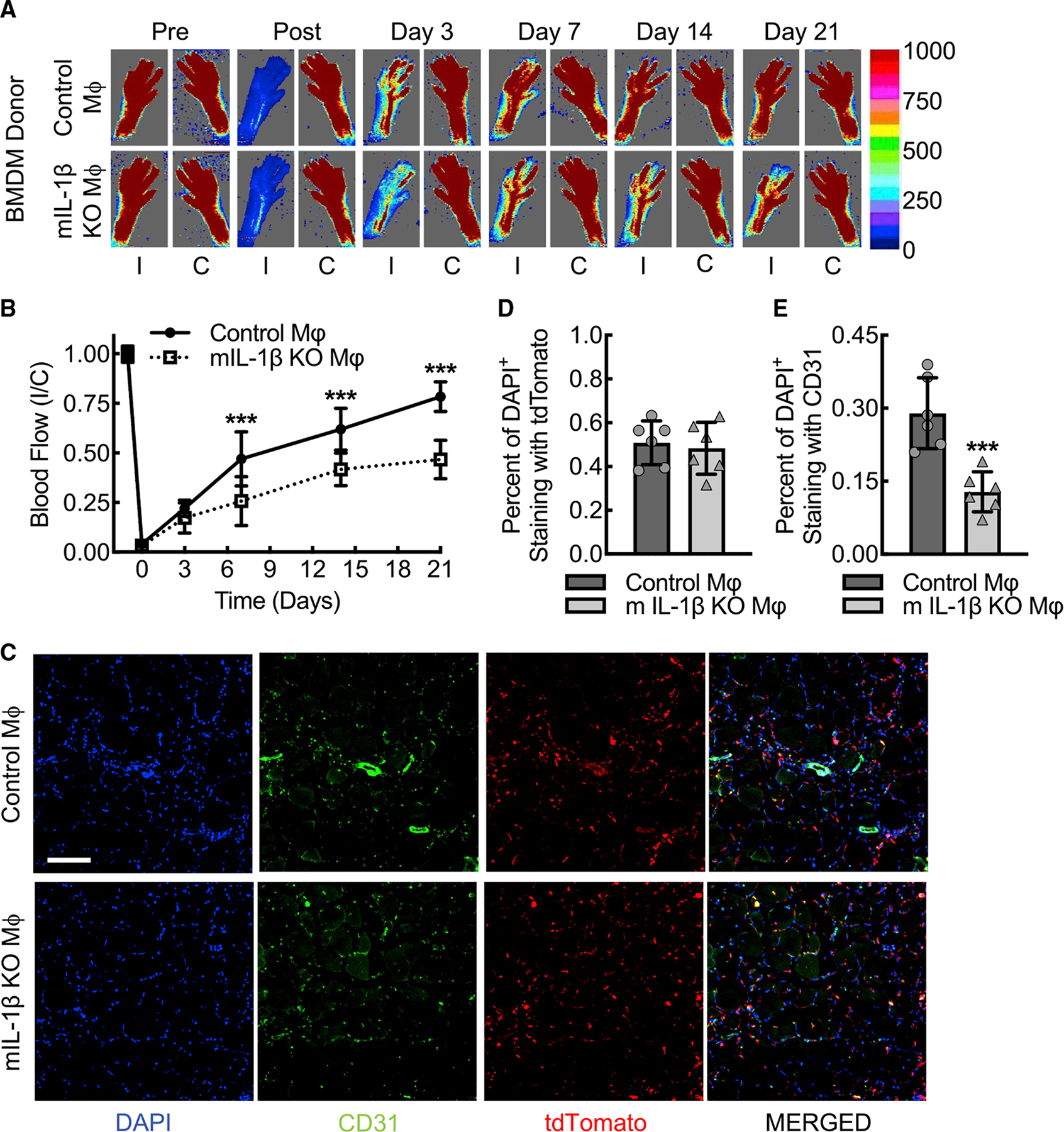
(A and B) Laser Doppler images of flow in the ischemic (I) and contralateral control (C) hindlimbs from myeloid IL-1β-deleted mice (mIL-1β KO) that underwent clodronate liposome macrophage depletion followed by transplant of tdTomato-labeled BMDMs from either wild-type (Control Mφ) or IL-1β-deleted (mIL-1β KO Mφ) Ai9 mice along with quantitative analysis (B) (***, p ≤ 0.0003 compared between control Mφ and mIL-1β KO Mφ for each time point by ANOVA; n = 6 mice total, three males and three females).
(C–E) Immunofluorescence micrographs of ischemic muscle tissue at day 3 post femoral artery ligation mice treated as in (A) along with quantitation of DAPI+tdTomato+ (D) and DAPI+CD31+ (E) cells (***, p = 0.0008 by t test; n = 6 mice, three males and three females). Bar, 100 microns. Data, mean ± SD.