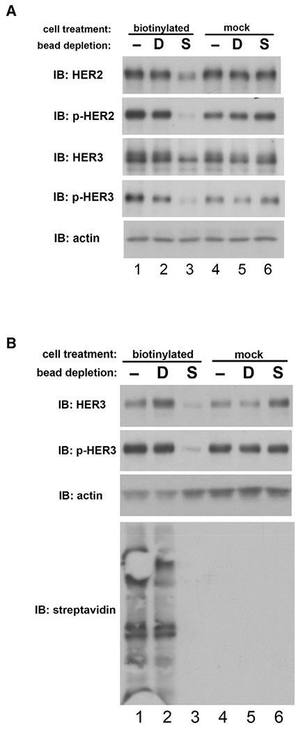
Figure 6. Constitutive HER3 signaling occurs almost entirely from the plasma membrane, not the intracellular pools.

(A) The surface of CHO cells expressing HER2 and HER3 was biotinylated using a cell-impermeable reagent. The entire surface proteome was then depleted from cell lysates using streptavidin beads, and HER2-HER3 expression and signaling activity was assayed in the intracellular lysate as shown. Lane 3 shows the membrane-depleted intracellular lysate; all other lanes are various negative controls. S indicates depletion by streptavidin beads, D indicates dummy beads, and – indicates no beads.
(B) The same experiment was performed on HER2-amplified HCC1569 breast cancer cells. HER3 signaling in the intracellular lysate was assayed as shown. The streptavidin immunoblot shows the total depletion of the surface proteome in experimental lane 3.
