
Figure 10.
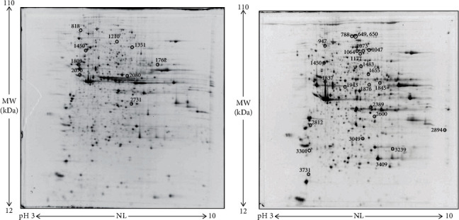
Representative 2DE images of protein extracts of bodies of both female (a) and male (b) flies supplemented with 100 μM PTS for 15 days. Protein extracts were separated in a 3–10 nonlinear gradient. SDS-PAGE was performed using 12% acrylamide. Gels were stained with ruthenium. Spot numbers indicate all the proteins identified by nLC-ESI-MS/MS and refer to the number reported in Table 2.