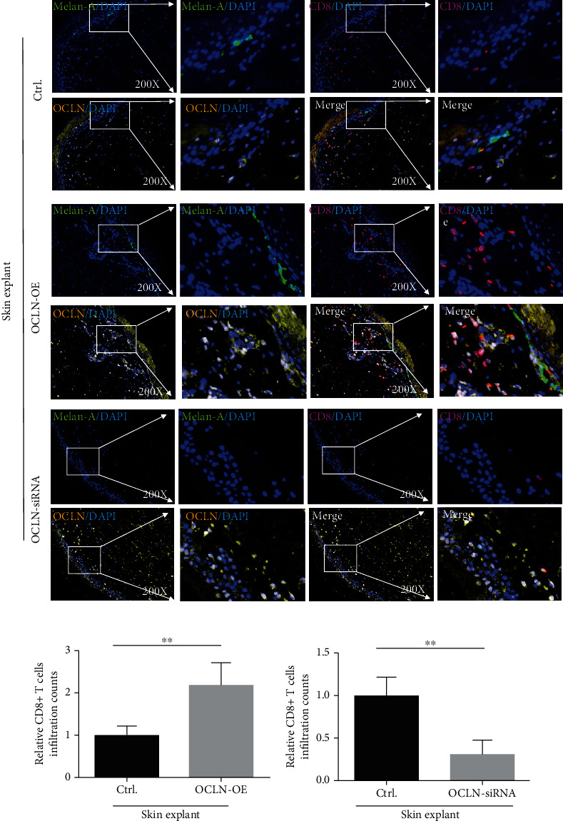
Figure 3.

The skin infiltration of CD8+ T cells in skin explants (n = 4, ∗∗P < 0.01, ∗∗P < 0.01). The red mark represents CD8-positive staining, the green mark represents Melan-A-positive staining (a melanocyte-specific antibody), the blue mark represents DAPI, and the yellow mark and white mark represent OCLN-positive staining, because when yellow light and blue light overlap, it is displayed as white.
