Figure 4.

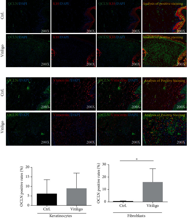
(a, b) The protein expression level of OCLN in keratinocytes from vitiligo patients and normal control (n = 5, P > 0.05). The red mark represents K10-positive staining, the green mark represents OCLN-positive staining, and the blue mark represents DAPI. The yellow mark of images analysis of positive staining represents the part where K10 and OCLN are colocalized after analysis by Inform2.3 software. (c, d) The protein expression level of OCLN in fibroblasts from vitiligo patients and normal control (n = 5, ∗P < 0.05). The red mark represents vimentin-positive staining, the green mark represents OCLN-positive staining, and the blue mark represents DAPI. The yellow mark of image analysis of positive staining represents the part where vimentin and OCLN are colocalized after analysis by Inform2.3 software.
