Figure 1.

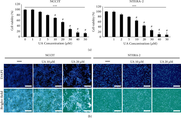
The UA has blocked the proliferation of embryonic CSCs. (a) MTT results showed inhibition of NTERA-2 and NCCIT cell proliferation after treatment with increased UA concentration for 24 hours. The data represent three independent tests. #p < 0.001 versus control. ∗∗∗p < 0.001 (ANOVA test). (b) UA created a nuclear deterioration in the embryonic CSCs. Class comparison microscopy images showing abnormal nucleus formation caused by 24-hour treatment at UA of 10 or 20 µM in NTERA-2 and NCCIT cells. Representing images are displayed (scale bar: 200 μm).
