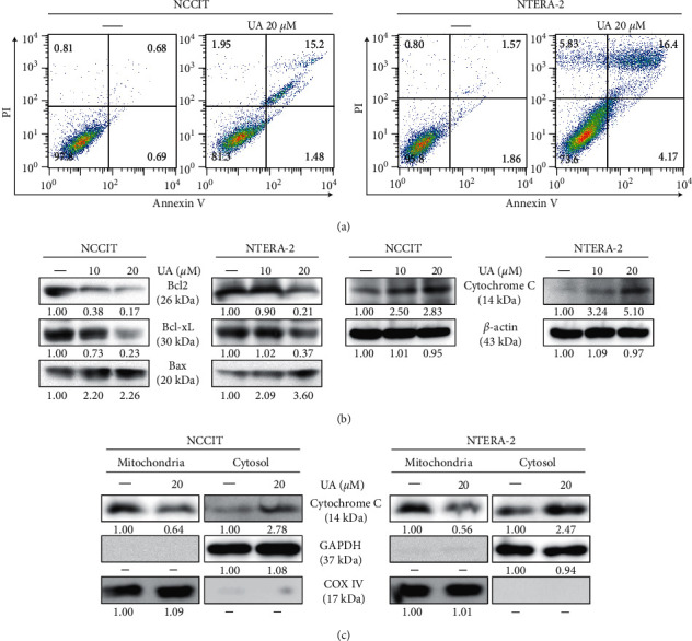
Figure 5.

UA induces the intrinsic apoptotic pathway. (a) Fluorescein-conjugated annexin V (annexin V-FITC) versus propidium iodide (PI) staining analysis in NTERA-2 and NCCIT cells showed apoptosis induction after treatment with 20 μM UA for 24 h. (b) Immunoblotting of NTERA-2 and NCCIT cells treated with 10 or 20 μM UA for 24 h showed levels of BCL-2, cytochrome. (c) BCL-xL and BAX expression. Exposure rates were measured by densitometry and were standardized in β-actin levels. Data were obtained in triplicate. Immunoblotting of cytochrome protein c in cytosolic and mitochondrial fractions separated from NTERA-2 and NCCIT cells after 24-hour treatment at 20 µM UA. Rate levels of cytosolic cytochrome c were calculated by densitometry and are normalized in GAPDH, and levels of mitochondrial cytochrome c were calculated by densitometry and were standardized in COXIV.
