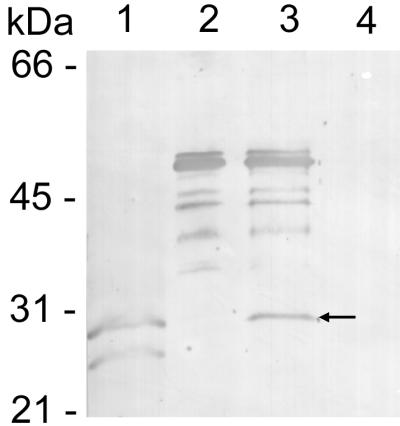
FIG. 2.

Protein immunoblotting of MAb 1A9 reacted with the p28 recombinant protein. Lanes: 1, heat-denatured E. chaffeensis (Arkansas strain) antigen; 2, GST fusion protein with p28; 3, thrombin-cleaved GST fusion protein (the arrow indicates the thrombin-cleaved recombinant p28); 4, GST protein only. The multiple bands in lanes 2 and 3 were apparently degradation products of the GST fusion protein.
