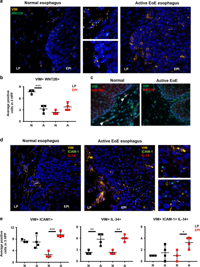
Fig. 4. Localization of VIM + cells expressing homeostatic and inflammatory markers reveals functional changes in EoE.
a Representative images of normal (n = 3) and active EoE esophagus (n = 4) hybridized with specific probes for VIM (yellow) and WNT2B (green, LP = lamina propria, EPI = epithelium). b Immunofluorescence staining of normal (n = 3) and active EoE esophagus (n = 4) for VIM (green) and WNT2B (red). c Quantification of VIM+ WNT2B+ cells in LP and EPI comparing normal and active EoE esophagus. d Representative images of normal (n = 3) and active EoE esophagus (n = 4) hybridized with specific probes for VIM (yellow), ICAM-1 (green) and IL-34 (red). e Quantification of VIM+ ICAM-1+ , VIM+ IL-34+ and VIM+ ICAM-1+ IL-34+ cells in LP and EPI comparing normal and active EoE esophagus. White arrows in c point positive cells. *p < 0.05, **p < 0.01 and ***p < 0.001.

