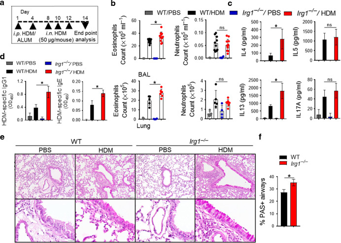
Fig. 2. Th2 cytokine production and allergic sensitization are increased in HDM-challenged Irg1−/− mice.
HDM was administered to WT (black) or Irg1−/− (red) mice. a The diagram shows the HDM administration (i.p., intraperitoneal; i.n., intranasal) and analysis schedule. b Scatter plot with bar show the numbers of CD11b+ Siglec-F+ eosinophils and CD11b+ Ly-6G+ neutrophils in BAL (top) and lung (bottom). c Bar charts show the cytokine secretion by ex vivo cultures of MedLN cells that had been re-stimulated with HDM (100 μg ml−1), d Serum HDM-specific IgE and IgG1 levels. e Images show histopathological sections of HDM-challenged lung from WT or Irg1−/− stained with PAS. Scale bars, 100 μm for ×100 and 50 μm for ×400 images. f Enumerated PAS+ airways from histopathological lung sections (n = 5–7 mice, one of 3 representative experiments is shown; *P < 0.05, WT versus Irg1−/−, unpaired t-test, two independent experiments).

