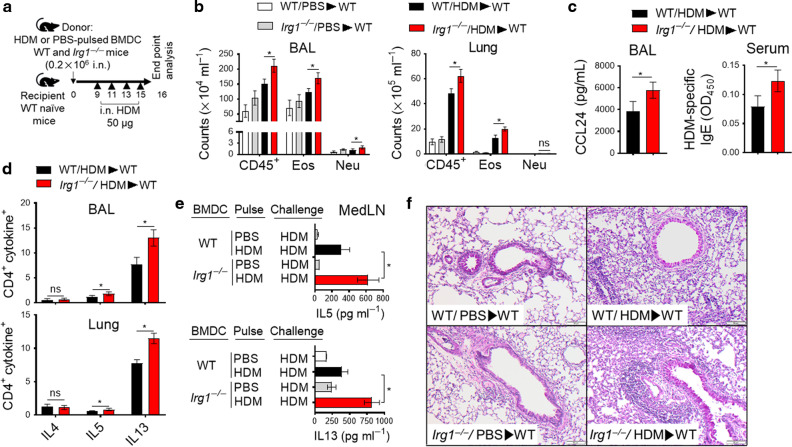
Fig. 3. Adoptive transfer of BMDC from Irg1−/− mice promotes HDM-mediated type 2 airway inflammation in WT mice.
a BMDC from WT or Irg1−/− mice were HDM-pulsed (100 μg/ ml) or sham-pulsed with PBS for 16 h and were adoptively transferred to naïve WT mice (0.2 × 10⁶ cells in 40 μl sterile saline; i.n. administration). Recipient WT mice received HDM challenges (50 μg; i.n) on days 9, 11, 13, and 15 before harvest on day 16. b The number of total CD45+ inflammatory cells, eosinophils, and neutrophils in recipient WT BAL and lung; c BAL CCL24 (left) and serum levels of HDM-specific IgE (right) and d quantification of Th2 immune response, as assessed based on the percentage of CD4+cytokinev+ cells in BAL and lung. e IL5 and IL13 cytokine secretion in culture supernatants of MedLN cells were re-stimulated with or without HDM (100 μg/ml). f Representative histopathological sections of PAS-stained WT recipient lungs. Scale bars, 100 μm for ×100. Data are expressed as mean ± SEM, and one of the three adoptive transfer experiments is shown; (n = 4–5 mice per group, *P < 0.05, WT/HDM/WT versus Irg1−/−/ HDM/ WT, unpaired t-test).

