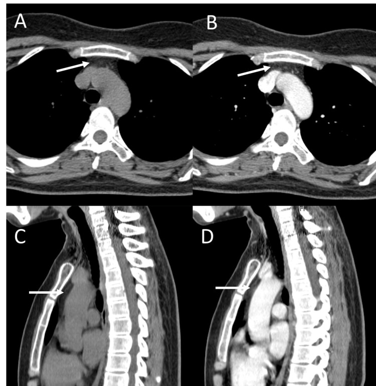
Figure 1. MDCT of the chest.
(A) Axial non-contrast, (B) axial with contrast, (C) sagittal non-contrast, and (D) sagittal with contrast views show prominent soft tissue without abnormal enhancement at mid-superior mediastinum in the prevascular space anterior to the great vessels (arrow).
MDCT, Multidetector computed tomography.

