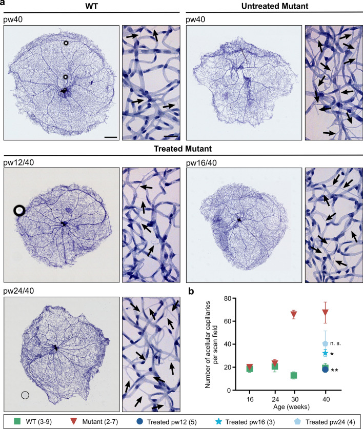
Fig. 7.
Quantification of acellular capillaries. Untreated Pde6bSTOP/STOP (mutant) and Pde6bSTOP/+ (WT) mice were analyzed at 16, 24, 30, and 40 weeks of age. Pde6bSTOP/STOP mutant mice were treated at 12, 16 or 24 weeks of age, and all analyzed at 40 weeks of age. Whole-mounted retinas were trypsin digested and stained with hematoxylin–eosin. a Representative images of whole retinal vasculature and higher-magnification images—all at 40 weeks of age. Black arrows, acellular capillaries; scale bars, 500 µm (whole retina/low mag) and 25 µm (high mag). b Mean number of acellular capillaries per scan field (± SEM); t test comparing treated mutants vs 40-week-old untreated mutant; n.s. not significant; *P ≤ .05; ** P ≤ .01. N values are provided in the legend next to the groups and in detail in “Materials and methods”

