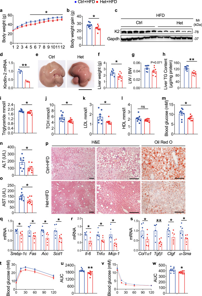
Fig. 2. Kindlin-2 haploinsufficiency ameliorates HFD-induced hepatic steatosis.
a Bodyweight. Six-week-old control (Ctrl: Cre-negative Kindlin-2fl/fl) and Kindlin-2 Het male mice (Het: Alb-Cre; Kindlin-2fl/+) were fed with HFD for 12 weeks (n = 8). b Body weight gain after HFD for 12 weeks (n = 8). c Western blotting. The kindlin-2 expression after HFD feeding was examined by western blotting. Protein extracts (20 μg) were used for western blotting from each sample (n = 6). d qRT-PCR analysis. Kindlin-2 mRNA expression after HFD feeding was examined by qRT-PCR analyses (n = 3). e Gross liver appearance. Scale bar, 1 cm. f Liver weight (n = 8). g Liver body ratio was measured (n = 8). h Liver TG content (n = 8). i–m Serum TG, TCH, LDL, HDL, and glucose levels (n = 8). n, o Serum ALT and AST levels (n = 8). p Representative H/E staining and Oil Red O staining of liver sections. Scale bar, 100 μm. q–s qRT-PCR analyses. mRNA expression levels of the indicated genes in liver samples from control/HFD and Het/HFD groups were determined (n = 6). t Glucose tolerance tests (GTT). Six-week-old male mice fed on HFD for 12 weeks were subjected to GTT. u Area under the curve (AUC) calculated based on s (n = 8). v Insulin tolerance tests (ITT). Six-week-old male mice fed on HFD for 12 weeks were subjected to ITT. w AUC calculated based on u (n = 8). a, b, d, f–o, q–s, u, w Data are presented as mean ± SEM. *P < 0.05, **P < 0.01, determined by two-tailed Student’s t-test. p Data are representative of three biologically independent replicates.

