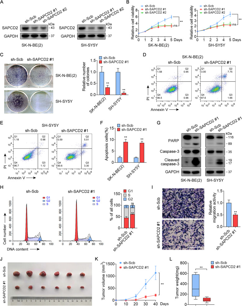
Fig. 2. SAPCD2 sustains NB cells growth and survival in vitro and in vivo.
A Western blot revealing SAPCD2 expression in NB cells stably transfected with scrambled shRNA (sh-Scb) or sh-SAPCD2. B CCK-8 assay indicates the change in cell viability of NB cells transfected with sh-Scb or sh-SAPCD2. C Representative images and quantification of colony formation assay depicting the growth of NB cells transfected with sh-Scb or sh-SAPCD2. D–F Flow cytometry showing the apoptosis of NB cells transfected with sh-Scb or sh-SAPCD2. G Western blot revealing caspase-3 activation and cleavage of PARP in NB cells transfected with sh-Scb or sh-SAPCD2. H Flow cytometry showing cell cycle distribution of SK-N-BE(2) cells transfected with sh-Scb or sh-SAPCD2. I Representative images and quantification of transwell assay showing the cell migration of SK-N-BE(2) cells transfected with sh-Scb or sh-SAPCD2. Scale bars, 200 μm. J–L Representative images, in vivo growth curves and tumor weight at the endpoints of subcutaneous xenografts of SK-N-BE(2) cells transfected with sh-Scb or sh-SAPCD2 into the dorsal flanks of BALB/c nude mice (n = 6 per group). One-way ANOVA with Bonferroni’s multiple comparison test for analysis in B and K; unpaired two-sided t-test in C, F, H, I, and L, data were shown as mean ± SD (error bars). ns, not significant. Data were representative of three independent experiments.

