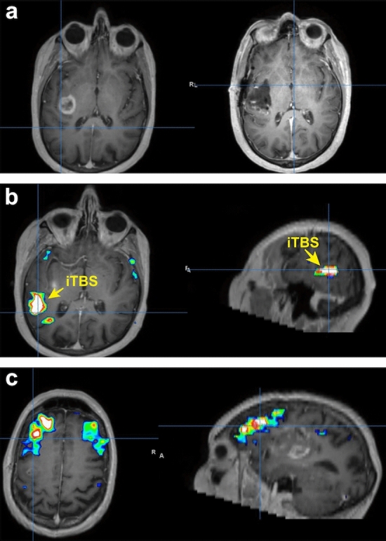
Figure 6.

TMS strategy for patient presenting with moderate expressive aphasia secondary to glioblastoma. (a) Preoperative MRI (left) demonstrating left insula glioblastoma and postoperative MRI (right) demonstrating complete resection. (b) Network analysis demonstrating a strongly organized posterior temporal region that is not in in the same network as Broca’s area. This was the area that was selected for treatment with iTBS. (c). Further network analysis demonstrating Broca’s area with bilateral representation that is not in the same network as the posterior temporal region.
