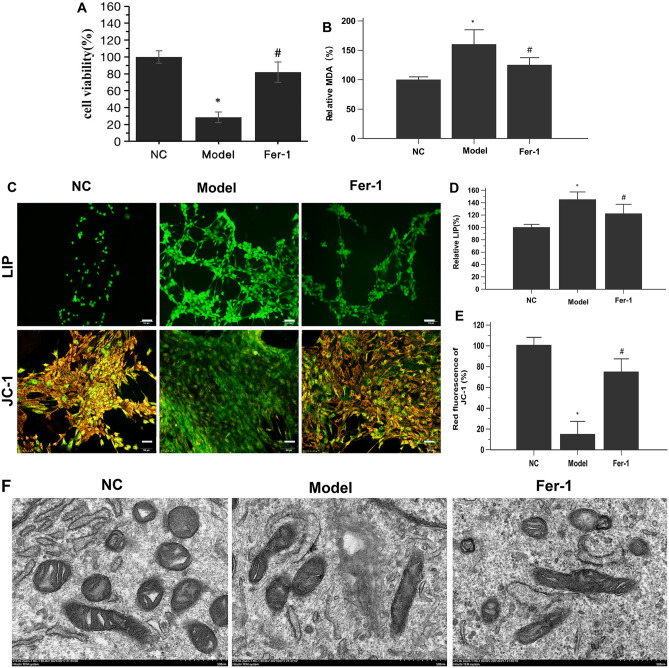
Figure 1.
Ferroptosis occurs in PAECs derived from MCT-induced PH rats. (A) Fer-1 restored the viability of MCT-induced PH rat-derived PAECs. (B) Fer-1 reduced lipid peroxidation in MCT-induced PH rat-derived PAECs. (C) Representative LIP and JC-1-stained PAECs. Morphology was examined using light microscopy. Scale bar = 100 μm. (D) Quantification of the relative LIP value in the different groups of PAECs. (E) Quantification of the relative value of JC-1 red fluorescence in the different groups of PAECs. (F) Representative TEM images of PAECs in the different groups show mitochondrial morphology. PAECs in the model group showed mitochondrial damage with mitochondrial outer membrane rupture and mitochondrial crista disappearance. Fer-1 restored these changes. Scale bar = 500 nm. NC, normal control group; Model, model group; Fer-1, MCT + Fer-1 (10 nM, treated for 4 h) group. Each experiment was repeated 3 times, n = 3. The data represent the means ± SD. *P < 0.05 versus the NC group, #P < 0.05 versus the model group.

