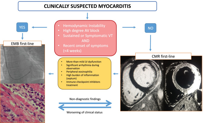
Fig. 2.
Proposed diagnostic algorithm in patients with suspected acute myocarditis. The figure shows when an endomyocardial biopsy (EMB) should be the first-line exam and when cardiac magnetic resonance (CMR) should be the first-line exam. The images on the left show a fluoroscopic-guided biopsy, with evidence on histology of eosinophilic myocarditis remaking the unique value of biopsy that can determine the type of inflammatory infiltrate. On the right, CMR images show the extent of late gadolinium enhancement and edema on T2-weight short tau inversion recovery sequences. The advantage of CMR is providing a complete vision of the heart showing the extent and the localization of the inflammatory process. AV indicates atrioventricular; VT, ventricular tachycardia; LV, left ventricular

