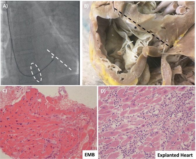
Fig. 4.
Right ventricular endomyocardial biopsy (EMB). A Fluoroscopy-guided biopsy from the right internal jugular vein. The bioptome passes the tricuspid valve (dashed oval) and reaches the ventricular septum (* and dashed line). B Explanted heart seen from the right ventricle of a patient with lymphocytic fulminant myocarditis shows the relationship between tricuspid valve and the ventricular septum (* and dashed line). Tricuspid chordal rupture can be a complication of biopsy from the right jugular vein. C From the same patient, the result of hematoxylin–eosin stain did not show an active myocarditis, while in D in the explanted heart an active lymphocytic myocarditis was demonstrated. This representative case underlines the false-negative results associated with biopsy due to the non-homogenous distribution of the inflammatory infiltrate and the potential sampling error

