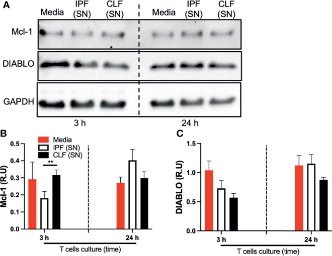
Figure 5.

The supernatant (SN) from IPF fibroblasts decreases Mcl-1 at the protein level in T cells at a short time of exposure. T cells obtained from four different healthy donors were stimulated with different IPF (SN) and CLF (SN) and recovered at the end of the culture. Then, Mcl-1 and DIABLO were examined by Western blot (A). Band densities were normalized with GAPDH by densitometry analysis and results are shown in relative units (RU) of Mcl-1 and DIABLO concentration (B, C, respectively) using ImageJ software. Bars indicate mean ± SD from four independent biological experiments. A multiple t-test and Holm–Sidak as post-test were used. **p < 0.01.
