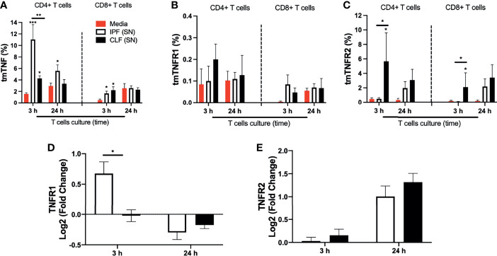
Figure 6.
The supernatant (SN) from IPF fibroblasts increases tmTNF expression and positively regulates TNFR1 at the transcriptional level in T cells at a short time of exposure. T cells obtained from five different healthy donors stimulated with different IPF (SN) and CLF (SN) were recovered at the end of the culture, and cells were prepared for flow cytometry and for obtention of RNA to perform a quantitative real-time PCR. The percentages of CD4+ T cells (left) and CD8+ T cells (right) positive to transmembrane (tm) TNF (A), tmTNFR1 (B), and tmTNFR2 (C) were obtained by flow cytometry. To acquire the transcriptional level, we calculated the log2 fold change of TNFR1 (D) and TNFR2 (E). Bars indicate mean ± SD from five independent biological experiments. A multiple t-test and Holm–Sidak as post-test (A–C) or a Mann–Whitney U test (D, E) was used. *p < 0.05, **p < 0.01, ***p < 0.001.

