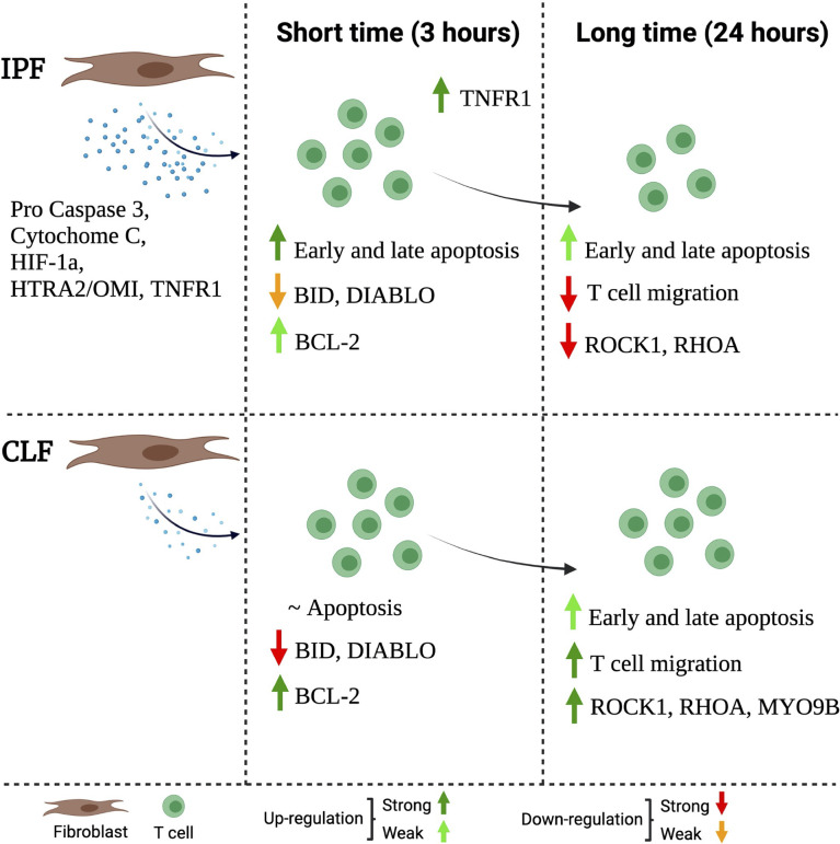
Figure 8.
Schematic representation of the proposed mechanism through IPF fibroblasts affects the function of T cells. The IPF fibroblast secretes high levels of pro-apoptotic molecules (left). This microenvironment, at a short time (3 h) of exposure, activates T-cell death, but at the same time, the T cells activate a weak anti-apoptotic profile as a defense mechanism. This profile is characterized by a slight loss of BID and DIABLO as well as a discrete presence of the Bcl-2; moreover, they increase TNFR1 expression (mild). At a long time of exposure (24 h), surviving T cells have decreased the expression of MYO9B, ROCK1, and RHOA, and consequently, these T cells are unable to migrate (right). The figure was created in BioRender.

