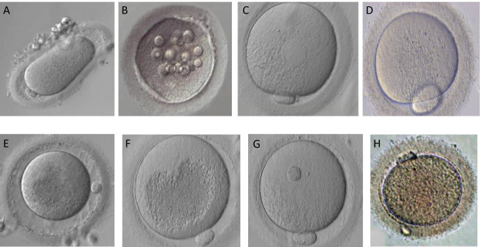
Fig. 2.
Different human oocyte morphological abnormalities (arrows) observed by light microscopy (400 × magnification): A, abnormal zona pellucida and cytoplasm shape; B, vacuoles; C, smooth endoplasmic reticulum clusters; D, giant polar body; E, large perivitelline space; F, centrally located cytoplasmic granulation; G, refractile body; H, brown oocyte

