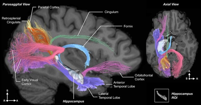
Figure 3 .

Example of the streamlines showing the hippocampal connections using diffusion tractography imaging for a single participant from the Human Connectome Project dataset. The tractography is from a transparent view through the brain, with parasagittal and axial slices of the human brain overlaid to show the trajectory of the pathways in the context of the brain. Voxels in the left hippocampus were seeded for the case illustrated. Streamlines from the hippocampus reach local areas such as the entorhinal and perirhinal cortex, and the parahippocampal gyrus (violet). However, in addition, streamlines reach more distant areas, including early visual cortical areas bilaterally via the (dorsal) hippocampal commissure (purple, pink), and the parietal cortex (yellow), the orbitofrontal cortex (magenta), the anterior cingulate cortex via the cingulum just above the corpus callosum (light green), and the anterior thalamus and mammillary bodies via the fornix (light blue). The hippocampus is shown in white with slight opacity and appears small posteriorly just because it rotates laterally (S, superior/inferior; L, left/right; A, anterior/posterior).
