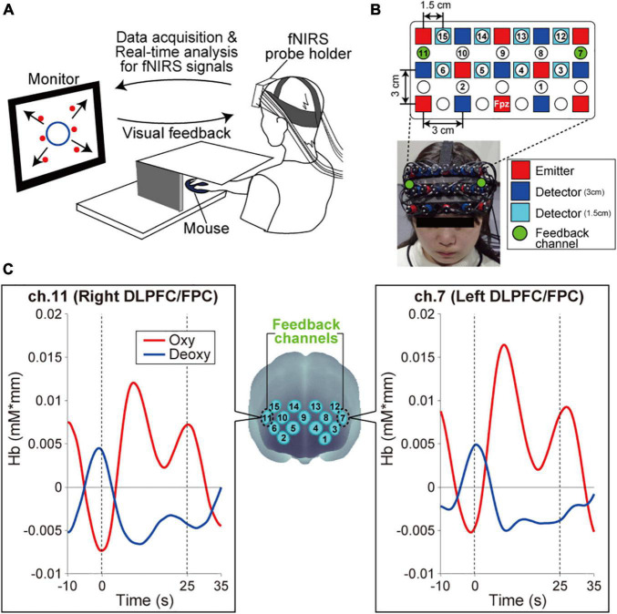
FIGURE 1.
Experimental setup. (A) Neurofeedback training set up. The monitor presented visual feedback on the status of participants’ bilateral prefrontal cortex in real-time. Furthermore, the six red circles on the monitor indicated the visual stimuli that participants were required to remember. Specifically, in each task block, the all-red circles were sequentially presented individually in random order at the predetermined fixed positions {with the center of the monitor as the origin, the coordinates of the six circles were [x(cm), y(cm)] = (–10, 8), (10, 8), (–10, 0), (10, 0), (–10, –8), and (10, –8)}. The appearance order changed with every task block. The participants were required to remember the spatial appearance order of six visual stimuli on the monitor. (B) Configuration of the fNIRS probe. Probes were placed over the prefrontal area. The channels, numbered 1–15, indicated the channels that output fNIRS signals that have been removed based on multidistance ICA. Ch.7 and ch.11 conveyed signals from the left and right DLPFC/FPC, respectively, as neurofeedback. (C) Spatial registration of the fNIRS maps onto MNI coordinate space. The left and right panels show typical profiles of oxy-Hb and deoxy-Hb in the feedback channels. Time zero indicates the onset of the task block. After starting the task block, the oxy-Hb signal showed a stronger response than the deoxy-Hb signal.

