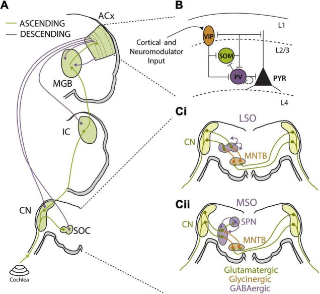
FIGURE 3.

Auditory circuit mechanisms for bottom-up and top-down adaptations. (A) Schematics of major auditory ascending (green) and descending (purple) pathways and associated auditory processing nuclei from the cochlea to auditory cortex (ACx). Major sound processing nuclei are highlighted in green, including the cochlear nucleus (CN), superior olive complex (SOC), inferior colliculus (IC), medial geniculate body (MGB), and ACx. Ascending pathways primarily terminate in layer 4 of ACx while corticofugal projections originate in layer 5b and layer 6 and can terminate at every level of the ascending pathway. (B) Summary figure showing known inhibitory relationships between parvalbumin (PV), somatostatin (SST), and vasoactive intestinal peptide (VIP)-positive neurons and their combined influences on excitatory cell populations. In this largely accepted model, locomotion, or top-down input, preferentially activates VIP cells, reducing SST cell output and releasing PV and excitatory cells from inhibition. PV activity can reduce background noise that improves signal-to-noise encoding of sensory stimuli by excitatory cell populations. Cortical layers are shown and separated by dashed lines. Figure was adapted from Pakan et al. (2016). (Ci,ii) Schematics of sound localization circuits of auditory brainstem used for processing interaural level (Ci) and timing (Cii) differences. (Ci) Principal neurons of lateral superior olive (LSO) receive excitatory glutamatergic inputs (green) from ipsilateral CN and glycinergic inhibition (orange) from ipsilateral medial nucleus for the trapezoid body (MNTB). MNTB receives excitatory input from contralateral CN. The interaction of ipsilateral excitation and contralaterally driven inhibition drives LSO firing in manner that can be used to calculate interaural level differences (ILD). LSO principal neurons release GABA (purple) in activity-dependent manner to asymmetrically modulate function of both glutamatergic and glycinergic inputs via activation of pre-synaptic GABAB receptors. (Cii) Principal neurons of the medial superior olive (MSO) receive excitatory glutamatergic inputs (green) from ipsilateral CN and contralaterally driven glycinergic inhibition (orange) from ipsilateral MNTB. MSO neurons also receive ipsilaterally driven inhibition from lateral nucleus of the trapezoid body (not shown). MSO neurons send excitatory projections to superior olivary nuclei (SPN), which in turn send feedback GABAergic projections (purple) to the MSO.
