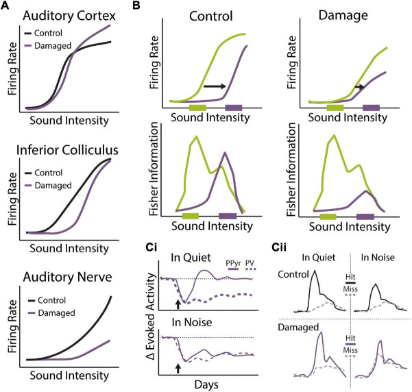
FIGURE 5.
Central gain enhancement following sensorineural hearing loss. (A) Schematics of rate-intensity functions from multiple levels of the auditory system under control conditions (black) or following cochlear damage via noise or ototoxic drug exposure that results in the central gain enhancement (purple). While output from the AN is severely degraded in terms of evoked-response threshold and suprathreshold intensity coding, rate-intensity functions gradually recover at ascending levels of the auditory system so that thresholds and suprathreshold responses are nearly normal at the level of the ACx and, in some cases, exhibit rebound hyperactivity. (B) Mean sound level adaptation to loud sound environments is altered with noise-induced hidden hearing loss. Rate-intensity functions from the IC of control (left) and noise exposed (right) mice when exposed to dynamic sound stimulus that switches between distributions with high probability of low sound levels (green) and high probability of high sound levels (purple). Noise exposed animals exhibit less dynamic range adaptation (top) and response functions carry less information about loud sound environments (bottom) compared to control animals. Schematized data adapted from Bakay et al. (2018). (Ci,ii) Cochlear degeneration triggers compensatory changes to cortical excitatory/inhibitory balance that differentially effects tone detection in quiet and noise. (Ci) Distinct changes to tone-evoked calcium transients in putative excitatory pyramidal neurons (PPyr, solid lines) and genetically-labeled PV inhibitory neurons (dashed lines) in the ACx following ouabain induced cochlear degeneration (arrow). Following transient loss of evoked activity, PPyr neurons exhibit near complete recovery of evoked-response size in quiet but not in background noise. Sustained decreases to tone-evoked activity in PV neurons are observed following ouabain treatment in both quiet and noise conditions. (Cii) Combined behavioral and imaging sessions showing differences in tone-evoked responses in cortical PPyr neurons on hit (solid lines) vs. miss (dashed lines) trials in quiet or background noise from animals before (control, black) and after ouabain-induced cochlear degeneration (damaged, purple). Decreased tone-in-noise detection in ouabain-treated animals is not only associated with diminished tone-evoked responses on hit trials but also increased activity on miss trials. These results suggest that altered E/I balance in the ACx following cochlear degeneration may lead to impaired adaption to background noise and decreased signal-to-noise ratios for detection of foreground stimuli. Schematized data adapted from Resnik and Polley (2021).

