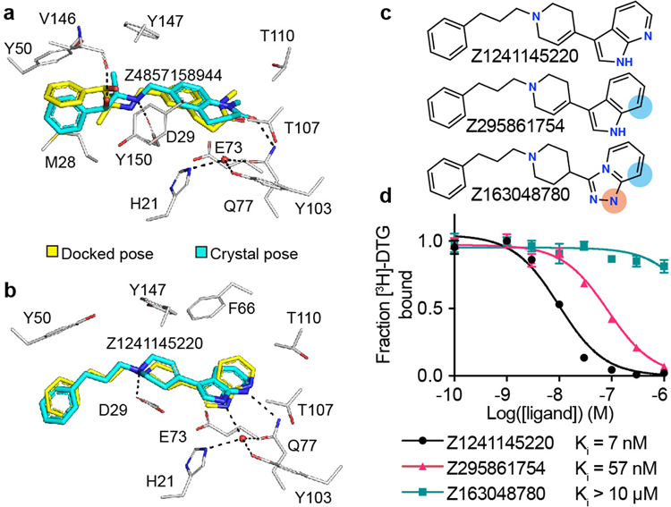
Figure 3 |. High structural fidelity between docked and crystallographic poses of novel σ2 receptor ligands.
Ligand crystal poses (carbons in cyan) overlaid with respective docked poses (yellow). σ2 receptor carbons are in grey, oxygens in red, nitrogens in blue, sulfurs in yellow, hydrogen bonds are shown as black dashed lines. a, Z4857158944-bound complex (PDB ID: 7M96; RMSD = 1.4 Å). b, Z1241145220-bound complex (PDB ID: 7M95; RMSD = 0.88 Å). c, Two Z1241145220 analogues that disrupt the hydrogen bonds with Gln77 and the structural water. Blue and apricot circles depict differences between the analogues and the parent compound. d, Competition binding curve of compounds from c. The data are the mean ± SEM from three technical replicates.

