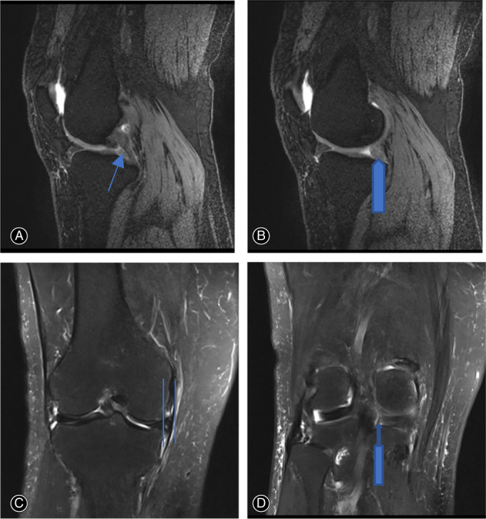
Fig. 1.

Preoperative MRI images of a 67‐year‐old woman patient with right knee MMPRT and normal limb alignment. (A) Ghost sign in T2 sagittal view indicated by long fine arrow (B) round shape (not as normal triangular shape) of medial meniscus posterior root in T2 sagittal view indicated by long coarse arrow (C) medial meniscus extrusion in T2 coronal view indicated by two lines (D) left sign in T2 coronal view indicated by long coarse upper arrow.
