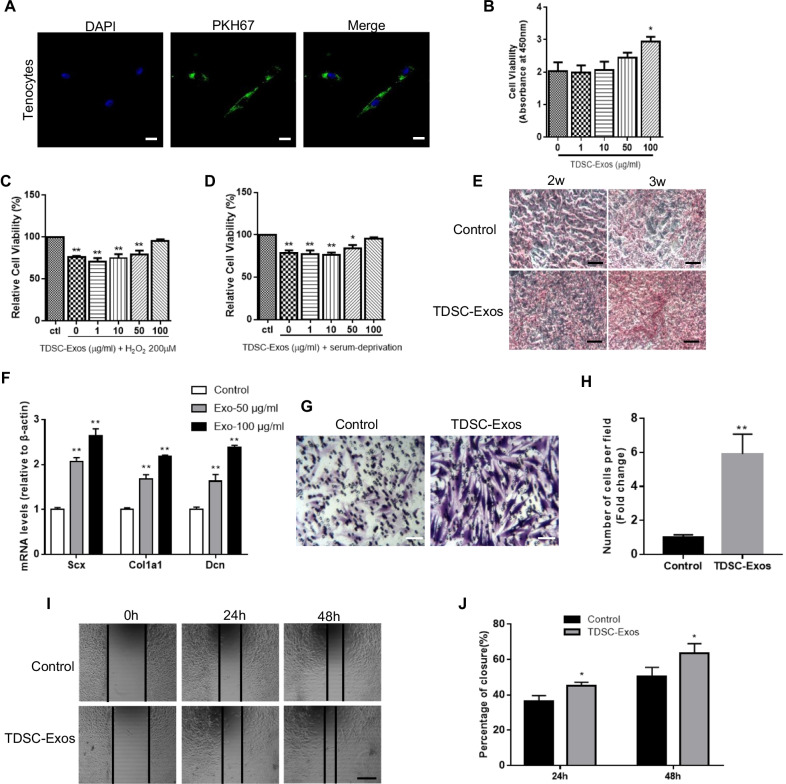
Fig. 2.
The effects of TDSC-Exos on tenocytes in vitro. A Tenocytes could absorb PKH67 (green)-labeled TDSC-Exos. The nuclei were stained with DAPI (blue). Scale bar: 20 μm. B Cell viability of tenocytes treated with TDSC-Exos at different concentrations (Bars: mean ± SE; n = 3; one-way ANOVA with Dunnett’s multiple comparison test, *p < 0.05 compared to control). C, D High concentration of exosomes could protect tenocytes from oxidative stress and serum deprivation (Bars: mean ± SE; n = 3; one-way ANOVA with Dunnett’s multiple comparison test, *p < 0.05 compared to control; **p < 0.01 compared to control). E Increased production of type I collagen was observed in tenocytes treated with TDSC-Exos by sirius red staining. Scale bar: 100 μm. F Tenocyte gene expression changes of Scx, Col1a1 and Dcn after treated with TDSC-Exos (Bars: mean ± SE; n = 3; one-way ANOVA with Dunnett’s multiple comparison test, **p < 0.01 compared to control). G Transwell assay of tenocytes treated with TDSC-Exos. Scale bar: 50 μm. H Quantitative analysis of the migrated cells in (G) (Bars: mean ± SE; n = 3; unpaired Student’s t test, **p < 0.01 compared to control). I TDSC-Exos promoted tenocyte migration in scratch wound healing assay. Scale bar: 250 μm. J Quantitative analysis about the percentage of closure in (I) (Bars: mean ± SE; n = 3; unpaired Student’s t test, *p < 0.05 compared to control)

