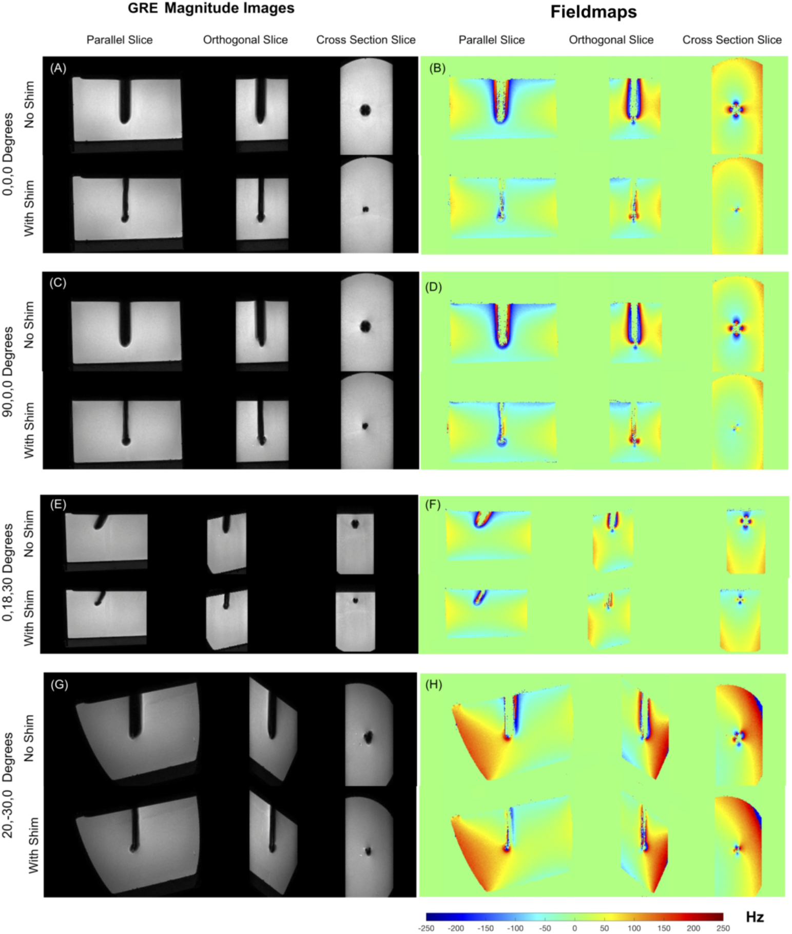
Figure 2.

Results of Needle shimming. (a-h) 1 x 1 x 1 mm3 3D GRE images and fieldmaps showing results of active shimming. Excellent recovery of lost signal is achieved in all orientations using pre-estimated shim currents. The width of the signal void approaches the needle width in all cases. Fieldmaps show correction of the underlying ΔB0. Without shimming, field wraps are observed closed to the needle due to extreme ΔB0 values, which is corrected with shimming. Also, regions closest to the needle with field information in the ‘Wth Shim’ case have no corresponding data in the ‘No Shim’ case due to the signal loss.
