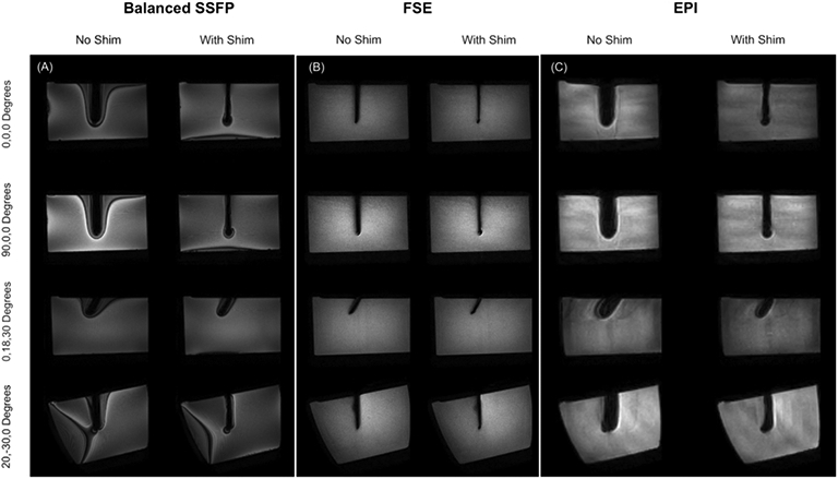
Figure 3.

Results of active shimming in 1 x 1 x 2 mm3 Balanced SSFP, 1 x 1 x 1 mm3 FSE and 2 x 2 x 2 mm3 3 shot EPI images. Recovery of lost signal is achieved in all sequences and all orientations. Balanced SSFP images show reduced off-resonance related banding. The void in the FSE images approaches the true width of the 4 mm needle after shimming.
