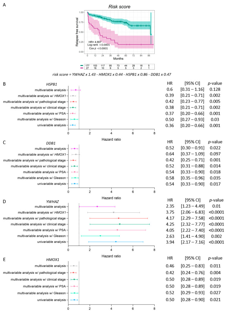
Figure 5.
Risk score regression model and multivariable analyses for HSPB1, DDB1, YWHAZ, and HMOX1 in PCa patients. (A) Kaplan–Meier curve for RFS in high-risk (purple lines) and low-risk (green lines) groups, according to a risk score model based on the expression of HSPB1, DDB1, YWHAZ, and HMOX1 in PCa patients. (B–D) Multivariable analyses presented by forest plots between each gene (HSPB1 (B), DDB1 (C), YWHAZ (D), and HMOX1 (E)) and GS, PSA, clinical and pathological stage, HMOX1′s expression, or all the variables together. Univariable analysis (light blue); multivariable analysis with GS (light green) = adjusted for the GS (6; 7 (3 + 4); 7 (4 + 3); 8-10); multivariable analysis with PSA (pink) = adjusted for the PSA serum levels at diagnosis (PSA (ng/mL): <4; 4-10; > 10); multivariable analysis with the clinical stage (dark green) = adjusted for the clinical stage; multivariable analysis with the pathological stage (red) = adjusted for the pathological stage; multivariable analysis with HMOX1 (grey) = adjusted for the expression of HMOX1; multivariable analysis (purple) = adjusted for all the variables simultaneously. HR = hazard ratios (95% confidence interval). All comparisons consider low expression patients as the reference group. Cox p = Cox proportional hazard model p-value. Statistical significance was set at Cox p < 0.05.

