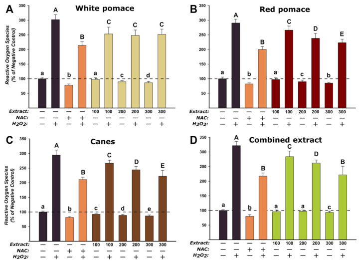
Figure 2.
Antioxidant effects of WPE (A), RPE (B), CE (C), and CoE (D) were evaluated using DCFH-DA assay on HGF. Cells were treated with the extracts (100, 200, and 300 µg/mL) or NAC (20 mM) for 24 h and further exposed to 50 µM DCFH-DA. The antioxidant potential was measured after a 2 h incubation in the presence and absence of 250 µM H2O2 (stimulated/non-stimulated conditions). The data are expressed as relative means ± standard deviation of three biological replicates, each one including 6 technical replicates. The values were expressed as relative values compared to the negative control (DMSO 0.2%) (100%). Different letters (a–e refers to comparisons in non-stimulated conditions, while A–E refers to comparisons in stimulated conditions) indicate statistically significant differences (ANOVA + Holm–Sidak post hoc test at p < 0.05).

